. 2022 Dec;103(6):234-244.
doi: 10.1111/iep.12456. Epub 2022 Oct 1.

Skin fibrosis associated with keloid, scleroderma and Jorge Lobo's disease (lacaziosis): An immuno-histochemical study

Affiliations

Skin fibrosis associated with keloid, scleroderma and Jorge Lobo's disease (lacaziosis): An immuno-histochemical study

Wagner Luiz Tafuri et al. Int J Exp Pathol. 2022 Dec.

Abstract

Fibrosis is a common pathophysiological response of many tissues and organs subjected to chronic injury. Despite the diverse aetiology of keloid, lacaziosis and localized scleroderma, the process of fibrosis is present in the pathogenesis of all of these three entities beyond other individual clinical and histological distinct characteristics. Fibrosis was studied in 20 samples each of these three chronic cutaneous inflammatory diseases. An immunohistochemical study was carried out to explore the presence of α-smooth muscle actin (α-SMA) and vimentin cytoskeleton antigens, CD31, CD34, Ki67, p16; CD105, CD163, CD206 and FOXP3 antigens; and the central fibrotic cytokine TGF-β. Higher expression of vimentin in comparison to α-SMA in all three lesion types was found. CD31- and CD34-positive blood vessel endothelial cells were observed throughout the reticular dermis. Ki67 expression was low and almost absent in scleroderma. p16-positive levels were higher than ki67 and observed in reticular dermis of keloidal collagen in keloids, in collagen bundles in scleroderma and in the external layers of the granulomas in lacaziosis. The presence of α-actin positive cells and rarely CD34 positive cells, observed primarily in keloids, may be related to higher p16 antigen expression, a measure of cell senescence. Low FOXP3 expression was observed in all lesion types. CD105-positive cells were mainly found in perivascular tissue in close contact with the adventitia in keloids and scleroderma, while, in lacaziosis, these cells were chiefly observed in conjunction with collagen deposition in the external granuloma layer. We did not find high involvement of CD163 or CD206-positive cells in the fibrotic process. TGF-β was notable only in keloid and lacaziosis lesions. In conclusion, we have suggested vimentin to be the main myofibroblast general marker of the fibrotic process in all three studied diseases, while endothelial-to-mesenchymal transition (EndoMT) and mesenchymal stem cells (MSCs) and M2 macrophages may not play an important role.

Keywords: fibrosis; immunohistochemistry; keloid; lacaziosis; scleroderma.


Conflict of interest statement

The authors declare no conflict of interest in this work.

Figures
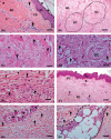
FIGURE 1
FIGURE 1
(A–H) Skin sections of patients with keloid, lacaziosis and scleroderma, (A) keloid showing a related rectification of the epidermal crests (asterisks). Note eosinophilic thickness collagen fibres (arrowheads) and a perivascular inflammatory cells exudate (arrows) (bar = 32 μm); (B) higher magnification of (A) showing large bundles of thick, hyaline and homogeneous collagen fibres called in the literature ‘keloid collagen’ (KC) (bar = 16 μm). (C) KC higher magnification of (A) showing spindle shaped cells (fibroblasts) (arrows) and eosinophilic thickness collagen fibres (arrowheads) (64 μm); (D) lacaziosis showing an inflammatory infiltrate comprising mononuclear cells and multinucleate giant cells (GC) (Langhans giant cell). Numerous yeast‐like cells of Lacazia loboi are present (arrows) (bar = 64 μm); (E) Large bundles of thick, homogeneous and hyaline collagen fibres (arrowheads) and spindle shape cells (fibroblasts) mixed to inflammatory cells as well (bar = 64 μm); (F) scleroderma showing thickening and homogenization of collagen bundles in the dermis. A focal chronic inflammatory reaction could be seen around skin appendages and blood vessels (arrowheads). (G) higher magnification showing this cellular exudate (arrowhead) and atrophical adnexal structures as eccrine glands (asterisks) (64 μm); (H) a typical straight edge of the dermal–subcutaneous interface was observed (dashed lines) (64 μm). Haematoxylin–eosin (HE) staining. Ep (Epithelium); Papillar Dermis (PD), Reticular Dermis (RD).
FIGURE 2
FIGURE 2
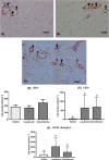
A–F: (A, B) a case of keloid to represent alpha‐Actin and vimentin immunohistochemical stanning, respectively; (A) higher magnification of a keloid nodule area showing endothelia vessel wall positive cells (arrows), but we can also observe some α‐SMA positive cells consisting of spindle‐shaped stromal cells (myofibroblasts) (arrowheads) in keloid collagen (KC) (reticular dermis) (16 μm); (B) compare vimentin expression to alpha‐Actin expression. Spindle‐shaped stromal cells (myofibroblasts) much more markedly visible than α‐Actin cell labelling throughout reticular dermis (16 μm). Immuno‐streptavidin–peroxidase method staining, Haematoxylin‐Harris counterstain. Figure C alfa‐Actin (α‐SMA) * a,b keloid versus lacaziosis (p = .0039); *a,c keloid versus scleroderma (p < .0001) and * b, lacaziosis versus scleroderma (P = .0664) Figure D vimentin * a,b keloid versus scleroderma (p < .001); a,b lacaziosis versus scleroderma (p < .001); b, keloid versus lacaziosis (p = .9905); * Figure E a case of keloid to represent TGF‐beta positive cells consisting of spindle‐shaped stromal cells (fibroblasts) (arrows) throughout the reticular dermis mixed to thick, homogenous and hyaline collagen fibers (asterisks) (16 μm); Figure F, keloid versus lacaziosis (p > .999); a,b keloid versus scleroderma (p < .0001); a,b lacaziosis versus scleroderma (p = .0007).
FIGURE 3
FIGURE 3
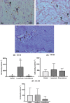
A–F: (A–C) a case of keloid to represent CD31, CD34, CD105, CD105 positive cells, respectively, localized along the blood vessels wall in the dermis (arrows). Except in a few cases as seen in (C), we observed some mesenchymal stromal cell population throughout the dermal compartment (arrowheads) (16μm). Immuno‐streptavidin–peroxidase method staining, Haematoxylin‐Harris counterstain. Figure D, CD31 Expression: * keloid versus lacaziosis (p > .9999); * keloid versus scleroderma (p > .9999); * lacaziosis versus x scleroderma (p = .08697). Figure E, CD34 Expression * a,b keloid versus lacaziosis (p > .9999); Figure F, CD105 Expression * a,b keloid versus lacaziosis (p = .0013); * a, keloid versus scleroderma (p = .9953); * b,c lacaziosis versus scleroderma (p = .0402).
FIGURE 4
FIGURE 4
A–F: (A–C) a case of keloid to represent CD68, CD63, CD206, respectively. (A,B) CD68 and CD163 positive cells, respectively keep a macrophage morphology and they were easily found occasionally distributed mixed composing the inflammatory cellular exudate (arrows). In (C), note CD206 positive cells was mainly distributed around blood vessels, but not strictly close to endothelium layer maintaining regularly distributed along external layers as well (arrowheads) (32 μm). Immuno‐streptavidin–peroxidase method staining, Haematoxylin‐Harris counterstain. Figure D. CD68 expression: * a,b lacaziosis versus keloid (p < .0001); a,b lacaziosis versus scleroderma (p < .0001); a, keloid and scleroderma cases (p = .9999).Figure E: CD163 expression: * keloid versus lacaziosis (p < .2452); * keloid versus (P < .7628); * lacaziosis versus (p < .6190). Figure F: CD206 expression: * keloid versus lacaziosis (p > .9999); *keloid versus scleroderma (p = .1275); * lacaziosis versus scleroderma (p = .0623).
FIGURE 5
FIGURE 5
A–F: (A,B) a case of scleroderma to represent KI67 and p16 immunohistochemical cellular characterization. (A) Any ki67 positive cells could be found in the dermis associated to fibroblasts (arrows) or to thick and homogenous collagen fibres (asterisks); (B) On the other side, many p16 positive cells throughout the fibrosis process; and homogenous collagen fibres (asterisks) as well (16 μm). (C) FOXP3 positive nuclei of cells consisting of spindle‐shaped stromal cells (fibroblasts) throughout the reticular dermis with homogenous collagen fibres (asterisks) could be seen (arrows) (16μm). Immuno‐streptavidin– peroxidase method staining, Haematoxylin‐Harris counterstain. Figure D Ki67 expression: a,b *keloid versus lacaziosis p < .9999. Figure E p16 expression: 17 a, *keloid versus lacaziosis p = .3112; a,b keloid versus scleroderma p = .0002; b, *lacaziosis versus scleroderma; p > .9999; Figure F Foxp3 expression * a,b keloid versus × lacaziosis p = .001; * a,b scleroderma versus lacaziosis p = .0071; * a, keloid versus scleroderma p > .9999.

Similar articles

Cited by

References

    1. Friedman SL. Mechanisms of disease: mechanisms of hepatic fibrosis and therapeutic implications. Nat Clin Pract. 2004;1:98‐105. - PubMed
    1. Wynn TA. Cellular and molecular mechanisms of fibrosis. J Pathol. 2008;214:199‐210. - PMC - PubMed
    1. Distler JHW, Györfi AH, Ramanujam M, Whitfield ML, Königshoff M, Lafyatis R. Shared and distinct mechanisms of fibrosis. Nat Rev Rheumatol. 2019;15:705‐730. - PubMed
    1. Taborda PR, Taborda VA, McGinnis MR. Lacazia loboi gen. Nov. comb. nov. the etiologic agent of lobomycosis. J Clin Microbiol. 1999;37:2031‐2033. - PMC - PubMed
    1. Ogawa R. Keloid and hypertrophic scars are the result of chronic inflammation in the reticular dermis. Int J Mo Sci. 2017;18:606‐616. - PMC - PubMed

Publication types