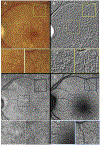
Enhanced Detection of Reticular Pseudodrusen on Color Fundus Photos by Image Embossing
- PMID: 36183241
- PMCID: PMC10350296
- DOI: 10.1080/02713683.2022.2126860
Enhanced Detection of Reticular Pseudodrusen on Color Fundus Photos by Image Embossing
Erratum in
-
Correction.Curr Eye Res. 2023 May;48(5):I. doi: 10.1080/02713683.2023.2200072. Epub 2023 Apr 11. Curr Eye Res. 2023. PMID: 37017369 No abstract available.
Abstract
Purpose: To evaluate whether processing a color fundus photo (CFP) using an image embossing technique can improve the detection of reticular pseudodrusen (RPD).
Methods: This post-hoc analysis included the eyes of subjects enrolled in the Amish Eye Study with early or intermediate age-related macular degeneration and evidence of RPD. All patients underwent CFP, near-infrared reflectance (NIR), and fundus autofluorescence (FAF) imaging. The ground-truth presence of RPD was established with a combination of NIR and FAF imaging. An embossing processed (EP) image was created by replacing each pixel of the CFP image with a highlight or a shadow representing light and dark boundaries in the original CFP image. The presence of RPD in CFP and EP images was assessed by two graders in a masked fashion and the sensitivity of CFP and EP for detection of RPD was evaluated. Cohen's kappa (k) was used to test inter-grader agreement for CFP and EP.
Results: A total of 106 eyes from 62 patients with RPDs were analyzed. The sensitivity for detection of RPD on CFP and EP was 63.2% (95%CI: 52.0%-74.4%) and 91.5% (95%CI: 85.0%-98.0%), respectively. The inter-rater reliabilities of CFP and EP for RPD detection were 0.81 and 0.84, respectively.
Conclusions: Embossing of CFP can improve the sensitivity for detection of RPD. The embossing technique can be a useful tool for better assessment of the true frequency of RPD in datasets where only CFP images are available.
Keywords: Age-related macular degeneration; embossing; image processing; reticular pseudodrusen.
Figures


References
-
- Wong WL, Su X, Li X, Cheung CM, Klein R, Cheng CY, Wong TY. Global prevalence of age-related macular degeneration and disease burden projection for 2020 and 2040: a systematic review and meta-analysis. Lancet Glob Health 2014;2(2):e106–16. - PubMed
-
- Spaide RF, Ooto S, Curcio CA. Subretinal drusenoid deposits AKA pseudodrusen. Surv Ophthalmol 2018;63(6):782–815. - PubMed
-
- Nassisi M, Lei J, Abdelfattah NS, Karamat A, Balasubramanian S, Fan W, Uji A, Marion KM, Baker K, Huang X and others. OCT Risk Factors for Development of Late Age-Related Macular Degeneration in the Fellow Eyes of Patients Enrolled in the HARBOR Study. Ophthalmology 2019;126(12):1667–1674. - PubMed
-
- Hogg RE, Silva R, Staurenghi G, Murphy G, Santos AR, Rosina C, Chakravarthy U. Clinical characteristics of reticular pseudodrusen in the fellow eye of patients with unilateral neovascular age-related macular degeneration. Ophthalmology 2014;121(9):1748–55. - PubMed
Publication types
MeSH terms
Grants and funding
LinkOut - more resources
Full Text Sources
Medical
Miscellaneous
