. 2022 Oct 2;79(10):537.
doi: 10.1007/s00018-022-04529-2.

Atypical induction of HIF-1α expression by pericellular Notch1 signaling suffices for the malignancy of glioblastoma multiforme cells

Affiliations

Atypical induction of HIF-1α expression by pericellular Notch1 signaling suffices for the malignancy of glioblastoma multiforme cells

Jungwhoi Lee et al. Cell Mol Life Sci. .

Abstract

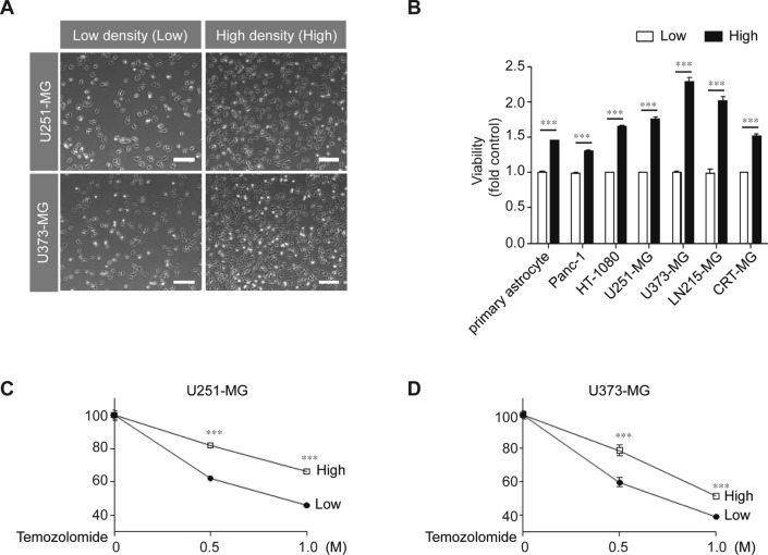
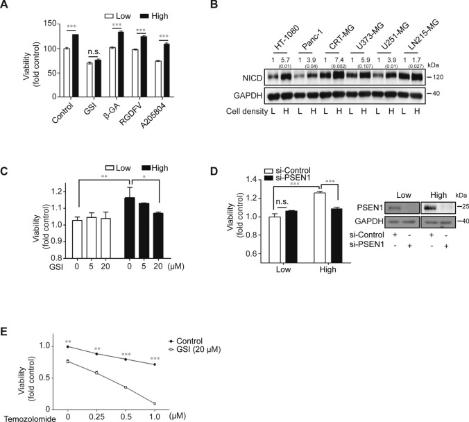
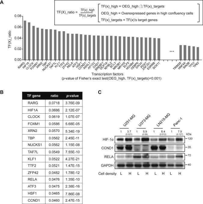
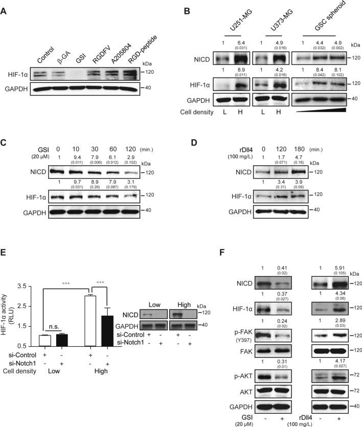
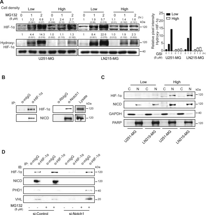
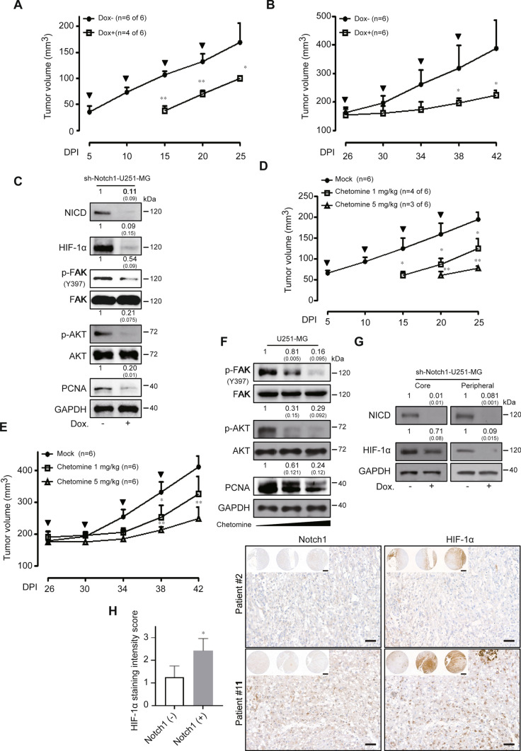
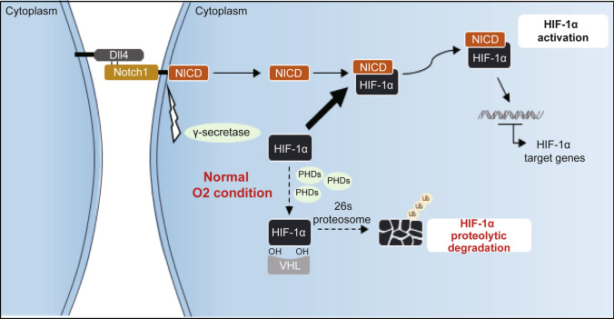
Contact-based pericellular interactions play important roles in cancer progression via juxtacrine signaling pathways. The present study revealed that hypoxia-inducible factor-1α (HIF-1α), induced even in non-hypoxic conditions by cell-to-cell contact, was a critical cue responsible for the malignant characteristics of glioblastoma multiforme (GBM) cells through Notch1 signaling. Densely cultured GBM cells showed enhanced viability and resistance to temozolomide (TMZ) compared to GBM cells at a low density. Ablating Notch1 signaling by a γ-secretase inhibitor or siRNA transfection resensitized resistant GBM cells to TMZ treatment and decreased their viability under dense culture conditions. The expression of HIF-1α was significantly elevated in highly dense GBM cells even under non-hypoxic conditions. Atypical HIF-1α expression was associated with the Notch1 signaling pathway in both GBM and glioblastoma stem cells (GSC). Proteasomal degradation of HIF-1α was prevented by binding with Notch1 intracellular domain (NICD), which translocated to the nuclei of GBM cells. Silencing Notch1 signaling using a doxycycline-inducible Notch1 RNA-interfering system or treatment with chetomin, a HIF pathway inhibitor, retarded tumor development with a significant anti-cancer effect in a murine U251-xenograft model. Using GBM patient tissue microarray analysis, a significant increase in HIF-1α expression was identified in the group with Notch1 expression compared to the group without Notch1 expression among those with positive HIF-1α expression. Collectively, these findings highlight the critical role of cell-to-cell contact-dependent signaling in GBM progression. They provide a rationale for targeting HIF-1α signaling even in a non-hypoxic microenvironment.

Keywords: Abnormal HIF-1α; Chemoresistance; GBM; Juxtacrine signaling; Notch1.


Conflict of interest statement

CC is the founder and shareholder of ILIAS Biologics, Inc. EK and KChoi are minor shareholders of ILIAS Biologics, Inc. The other authors declare that they have no competing interests.

Figures

Fig. 1
Fig. 1
High-density culture conditions induce malignant features in various cancer cells. A U251-MG and U373-MG cells were cultured at low (9.09 × 103 cells/cm2) or high density (2.38 × 104 cells/cm2). Scale bar indicates 100 µm. B Primary astrocytes and several cancer cell lines were cultured at low or high density for 24 h, after which, cell viability was measured using a WST-1 assay (n = 3; Tukey’s post hoc test was used to detect significant differences in ANOVA, p < 0.001; asterisks indicate a significant difference compared to Low density and High density, ***p < 0.001). C, D U251-MG and U373-MG cells cultured at low or high density were treated with 0.5 M or 1 M TMZ for 24 h, after which, cell viability was measured by the WST-1 assay (n = 3; Tukey’s post hoc test was applied to detect significant differences in ANOVA, p < 0.0001; asterisks indicate a significant difference compared to 0% inhibition, ***p < 0.001).
Fig. 2
Fig. 2
Ablating Notch1 signaling restrained density-dependent malignancy in various cancer cells. A U251-MG cells were treated with varying inhibitors associated with cell-to-cell interactions. GSI (50 μM), β-GA (50 μM), RGDFV (1 μM), or A205804 (30 μM) were administered for 24 h to low- or high-density cultured U251-MG cells, after which, cell viability was measured by the WST-1 assay (n = 3; Tukey’s post hoc test was used to detect significant differences in ANOVA, p < 0.001; asterisks indicate a significant difference compared to Low density and High density, ***p < 0.001). B The expression level of Notch intracellular domain (NICD) was measured by immunoblot assays in low- or high-density cultured cell lysates from HT-1080, Panc-1, CRT-MG, U373-MG, U251-MG, and LN215 cells. The relative pixel intensities were measured using ImageJ software. GAPDH was used as the loading control. The data are representative of three individual experiments. C U251-MG cells were treated with varying doses of GSI (0, 5, or 20 μM) for 24 h in low- or high-density cultured U251-MG cells, after which, cell viability was measured by the WST-1 assay (n = 3; Tukey’s post hoc test was applied to detect significant differences in ANOVA, p < 0.0001; asterisks indicate a significant difference compared to 0% inhibition, *p < 0.05, **p < 0.01). D Low- or high-density cultured U251-MG cells were transfected with control or PSEN1-targeting siRNA using Lipofectamine 2000™ reagent. After 48 h of siRNA transfection, cell viability was evaluated by the WST-1 assay (n = 3; Tukey’s post hoc test was used to detect significant differences in ANOVA, p < 0.001; asterisks indicate a significant difference compared to siControl and si-PSEN1, ***p < 0.001, n.s. non-significant). PSEN1 levels were significantly decreased in low- and high-density cultured U251-MG cells after si-PSEN1 transfections compared to si-Controls (Inset). E U251-MG cells were pre-treated with 20 μM of GSI. At 2 h post-treatment, the cells were incubated with or without varying doses of TMZ (0, 250, 500, and 1000 µM) for an additional 24 h, after which, cell viability was measured by the WST-1 assay (n = 3; Tukey’s post hoc test was applied to detect significant group effects in ANOVA, p < 0.0001; asterisks indicate a significant difference compared to 0% inhibition, *p < 0.05, **p < 0.01, ***p < 0.001)
Fig. 3
Fig. 3
Expression of HIF-1α is associated with cell density-dependent culture conditions. A Total mRNA was extracted from low- or high-density cultured U251-MG cells and analyzed by next-generation sequencing (NGS). Genes highly expressed in high-density cultured cells are indicated as OEG-high, target gene sets of transcription factors (x) are indicated as TF(x)_targets, and the intersection of the OEG-high gene set and each TF(x)_target is designated as the TF(x)_ratio. Each TF(x)_ratio of 199 transcription factors was calculated. B The top 15 ranked transcription factors are displayed with p values. C Expression levels of HIF-1α encoded by HIF1A gene, cyclin D1 encoded by CCND1 gene, and p65 (NF-κB p65 subunit) encoded by RELA gene in low- or high-density cultured U251-MG, U373-MG, LN215-MG, and Panc-1 cells were evaluated by immunoblot assays (L indicates low-density culture; H indicates high-density culture). GAPDH was used as the loading control. Relative pixel intensities were measured using ImageJ software. The data are representative of three individual experiments
Fig. 4
Fig. 4
Cell density-dependent HIF-1α expression is modulated by Notch1 signaling regulation. A β-GA, GSI, RGDFV-peptide, A205804, or RGD-peptide were administered to high-density cultured U251-MG cells, and HIF-1α expression was evaluated by immunoblot assays. GAPDH was used as the loading control. The data are representative of three individual experiments. B NICD or HIF-1α expression was measured at different confluencies of U251-MG and U373-MG cells by immunoblot assays. Glioblastoma stem cells (GSC) were cultured for 1, 4, or 7 days to establish different sizes of spheroids, after which, NICD or HIF-1α were measured by immunoblot assays. GAPDH was used as the loading control. Relative pixel intensities were measured using ImageJ software. The data are representative of three individual experiments. C, D GSI (20 µM) or recombinant Dll4 (rDll4; 100 mg/L) were added to U251-MG cells in a time-dependent manner, and then NICD or HIF-1α levels were analyzed by immunoblot assays. GAPDH was used as the loading control. Relative pixel intensities were measured using ImageJ software. The data are representative of three individual experiments. E Low- or high-density cultured U251-MG cells were transfected with control or Notch1-targeting siRNA using Lipofectamine 2000™ reagent. After 48 h of siRNA transfection, HIF-1α activity was measured by the luciferase reporter assay (n = 3; Tukey’s post hoc test was applied to detect significant group effects in ANOVA, p < 0.0001; asterisks indicate a significant difference compared to 0% inhibition, ***p < 0.001, n.s.: non-significant). NICD levels were significantly decreased in low and high-density cultured U251-MG cells after si-Notch1 transfections compared to si-Controls (Inset). F Low- or high-density cultured U251-MG cells were maintained for 24 h, after which, the expression levels of NICD, HIF-1α, p-FAK(Y397), FAK, p-AKT, AKT, or GAPDH were evaluated by immunoblot assays. Relative pixel intensities were measured using ImageJ software. The data are representative of three individual experiments
Fig. 5
Fig. 5
Interaction between NICD and HIF-1α prevents HIF-1α hydroxylation and degradation. A Low- or high-density cultured U251-MG and LN215-MG cells were treated with 5 μM of the proteasome inhibitor MG132 for different times (0, 1, or 2 h). In each cell lysate, HIF-1α or hydroxylated HIF-1α (hydroxy-HIF-1α) was evaluated by immunoblot assays. GAPDH was used as the loading control. Relative pixel intensities were measured using ImageJ software. The data are representative of three individual experiments. B High-density cultured U251-MG cell lysates were bound to anti-HIF-1α or normal IgG antibodies, followed by protein G agarose addition. The immunoprecipitated proteins were then detected by HIF-1α or NICD antibodies in immunoblot assays. Non-bound lysate was used for the loading control. C Low- or high-density cultured U251-MG and LN215-MG cells were separated into cytosolic and nuclear fractions, and HIF-1α or NICD levels were analyzed by immunoblot assays. GAPDH or PARP was used as the loading controls for the cytosolic and nuclear fractions, respectively. Relative pixel intensities were measured using ImageJ software. The data are representative of three individual experiments. D U251-MG cells were transfected with siControl or si-Notch1 for 48 h and treated with MG132 for 2 h before preparing cell lysates. Lysates immunoprecipitated with anti-HIF-1α or normal mouse IgG antibodies were immunoblotted using HIF-1α, NICD, PHD, or VHL antibodies. The data are representative of three individual experiments
Fig. 6
Fig. 6
Blockade of abnormal HIF-1α induces in vivo anti-cancer effects in GBM cells. A, B sh-Notch1-Dox (n = 6) and sh-Notch1-Dox+ (n = 6) U251-MG xenografted tumors were measured for 25 or 42 days. Bold arrows indicate the time of doxycycline treatment (Tukey’s post hoc test was used to detect significant differences in ANOVA, p < 0.0001; asterisks indicate a significant difference compared to sh-control and sh-Notch1, *p < 0.05, **p < 0.01, n.s. non-significant). C Western blot analysis of sh-Notch1-Dox and sh-Notch1-Dox+ tumor lysates using anti-Notch1, HIF-1α, p-FAK, FAK, p-AKT, AKT, and PCNA antibodies. GAPDH was used as the loading control. Relative pixel intensities were measured using ImageJ software. The data are representative of three individual experiments. D, E The effects of chetomin (0, 1, or 5 mg/kg) on tumor size in U251-MG xenograft models were analyzed after 25 and 42 days. The bold arrows indicate the time of chetomin injection (Tukey’s post hoc test has been used to determine significant group effects in ANOVA, P < 0.0001; asterisks indicate a significant difference between the control group and chetomin injection group, *P < 0.05). F Western blot analysis of 0, 1, or 5 mg/kg chetomin injection tumor lysates using anti-p-FAK, FAK, p-AKT, AKT, and PCNA antibodies. GAPDH was used as the loading control. Relative pixel intensities were measured using ImageJ software. The data are representative of three individual experiments. G Regional lysates (peripheral or core region) from sh-Notch1-Dox and sh-Notch1-Dox+ tumor samples were analyzed by immunoblotting for HIF-1α or NICD expression. GAPDH was used as the loading control. Relative pixel intensities were measured using ImageJ software. The data are representative of three individual experiments. Peripheral region refers to the outer 75% of the tumor sample sections. H Left, comparison of HIF-1α staining intensity scores between the Notch1-negative staining group (n = 4) and the Notch1-positive staining group (n = 7) among positively expressing HIF-1α patients. Right, representative images of GBM patient tissue microarray analysis (scale bar = 50 μm; inset image scale bar = 500 μm)
Fig. 7
Fig. 7
Graphical summary of non-hypoxic HIF-1α stabilization in GBM cells

Similar articles

Cited by

References

    1. Cloughesy TF, Cavenee WK, Mischel PS. Glioblastoma: from molecular pathology to targeted treatment. Annu Rev Pathol. 2014;9:1–25. doi: 10.1146/annurev-pathol-011110-130324. - DOI - PubMed
    1. Wolf KJ, Chen J, Coombes J, Aghi MK, Kumar S. Dissecting and rebuilding the glioblastoma microenvironment with engineered materials. Nat Rev Mater. 2019;4:651–668. doi: 10.1038/s41578-019-0135-y. - DOI - PMC - PubMed
    1. Meng X, Zhao Y, Han B, Zha C, Zhang Y, Li Z, Wu P, Qi T, Jiang C, Liu Y, Cai J. Dual functionalized brain-targeting nanoinhibitors restrain temozolomide-resistant glioma via attenuating EGFR and MET signaling pathways. Nat Commun. 2020;11:594. doi: 10.1038/s41467-019-14036-x. - DOI - PMC - PubMed
    1. Oshiro S, Tsugu H, Komatsu F, Ohmura T, Ohta M, Sakamoto S, Fukushima T, Inoue T. Efficacy of temozolomide treatment in patients with high-grade glioma. Anticancer Res. 2009;29:911–917. - PubMed
    1. Chang SM, Wen P, Cloughesy T, Greenberg H, Schiff D, Conrad C, Fink K, Robins HI, De Angelis L, Raizer J, Hess K, Aldape K, Lamborn KR, Kuhn J, Dancey J, Prados MD, C. North American Brain Tumor and I. the National Cancer Phase II study of CCI-779 in patients with recurrent glioblastoma multiforme. Investig New Drugs. 2005;23:357–361. doi: 10.1007/s10637-005-1444-0. - DOI - PubMed