. 2022 Oct 3;20(1):370.
doi: 10.1186/s12916-022-02571-2.

Probing SARS-CoV-2-positive plasma to identify potential factors correlating with mild COVID-19 in Ghana, West Africa

Affiliations

Probing SARS-CoV-2-positive plasma to identify potential factors correlating with mild COVID-19 in Ghana, West Africa

Kesego Tapela et al. BMC Med. .

Abstract

Background: West Africa has recorded a relatively higher proportion of asymptomatic coronavirus disease 2019 (COVID-19) cases than the rest of the world, and West Africa-specific host factors could play a role in this discrepancy. Here, we assessed the association between COVID-19 severity among Ghanaians with their immune profiles and ABO blood groups.

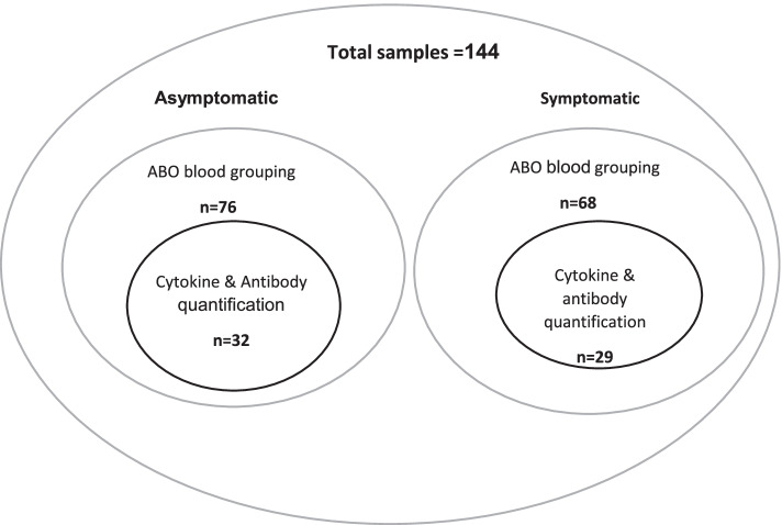
Methods: Plasma samples were obtained from Ghanaians PCR-confirmed severe acute respiratory syndrome coronavirus 2 (SARS-CoV-2)-positive individuals. The participants were categorized into symptomatic and asymptomatic cases. Cytokine profiling and antibody quantification were performed using Luminex™ multiplex assay whereas antigen-driven agglutination assay was used to assess the ABO blood groups. Immune profile levels between symptomatic and asymptomatic groups were compared using the two-tailed Mann-Whitney U test. Multiple comparisons of cytokine levels among and between days were tested using Kruskal-Wallis with Dunn's post hoc test. Correlations within ABO blood grouping (O's and non-O's) and between cytokines were determined using Spearman correlations. Logistic regression analysis was performed to assess the association of various cytokines with asymptomatic phenotype.

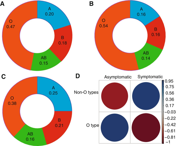
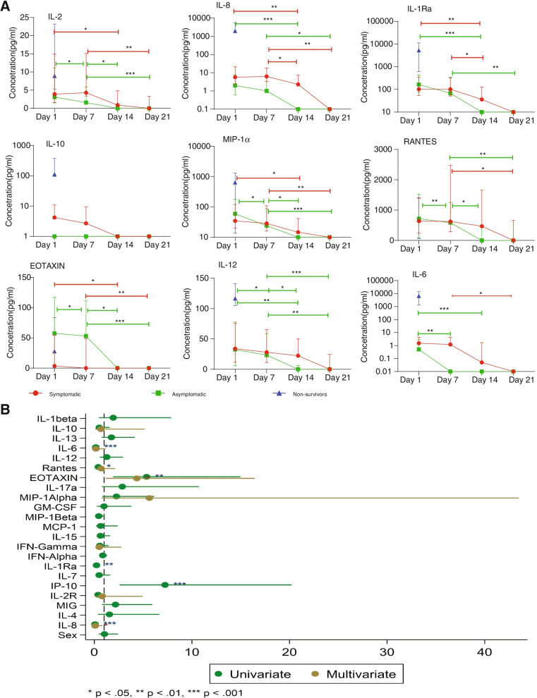
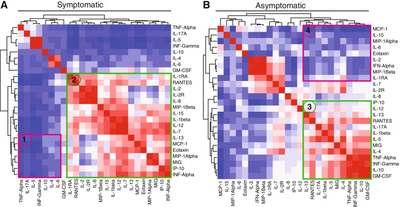
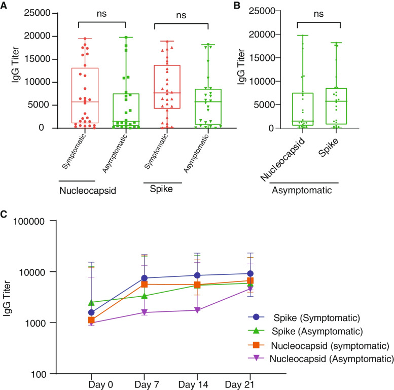
Results: There was a trend linking blood group O to reduced disease severity, but this association was not statistically significant. Generally, symptomatic patients displayed significantly (p < 0.05) higher cytokine levels compared to asymptomatic cases with exception of Eotaxin, which was positively associated with asymptomatic cases. There were also significant (p < 0.05) associations between other immune markers (IL-6, IL-8 and IL-1Ra) and disease severity. Cytokines' clustering patterns differ between symptomatic and asymptomatic cases. We observed a steady decrease in the concentration of most cytokines over time, while anti-SARS-CoV-2 antibody levels were stable for at least a month, regardless of the COVID-19 status.

Conclusions: The findings suggest that genetic background and pre-existing immune response patterns may in part shape the nature of the symptomatic response against COVID-19 in a West African population. This study offers clear directions to be explored further in larger studies.

Keywords: ABO blood groups; Antibodies; Asymptomatic; COVID-19; Eotaxin; West Africa.


Conflict of interest statement

The authors declare that they have no competing interests.

Figures

Fig. 1
Fig. 1
Sample distribution for different assays performed in the study
Fig. 2
Fig. 2
The distribution of blood group within asymptomatic and symptomatic and correlations between blood group and disease status. The distribution of the blood group is presented as ratios in a donut plot for all samples (A), asymptomatic (B) and symptomatic (C) and the correlations between blood group and disease status were presented as correlograms (D). The magnitude of Pearson residuals is shown on a colour scale of dark blue (positive correlation, 0.95–0.03) to white (no correlation) to red (negative correlation, −0.15 to −0.88) signal. The bigger the “dot”, the stronger or weaker the correlation coefficient
Fig. 3
Fig. 3
Cytokine concentration levels in asymptomatic and symptomatic cases. A Cytokine abundance among all COVID-19 patients and B comparison of cytokine concentration levels between COVID-19 symptomatic and asymptomatic patients. The cytokine concentration levels were measured from plasma of COVID-19 (n = 61) patients which were collected weekly for 4 weeks (medians of the time points). The bars depict the mean ± standard error of mean (SEM). The cytokine concentration levels were measured from plasma of COVID-19 symptomatic (n = 29) and asymptomatic (n =32) individuals. The median cytokine quantities per participant category obtained by extracting the medians across the time points per patient are shown by a horizontal line across the violin plots while the lower and upper dotted lines represent the 25th and 75th percentiles, respectively. Statistical significance between symptomatic and asymptomatic patients were determined by a two-tailed Mann-Whitney U test (ns: p >0.05, *p <0.05, **p < 0.01, ****p < 0.0001
Fig. 4
Fig. 4
Change in cytokine concentration levels over time and cytokine association in COVID-19-positive individuals. A The cytokine concentration levels were analysed from plasma of COVID-19 symptomatic (n = 29), asymptomatic (n = 29) and non-survivors (n = 2) patients at different timepoints for 4 weeks. The median quantity of the cytokines for each sampling day is shown by the line graph and the 25th and 75th percentiles are cytokine levels. Statistical significance between days was determined by Kruskal-Wallis (*p <0.05, **p < 0.01, ****p < 0.0001). B The association of the cytokines and antibodies to asymptomatic phenotype were estimated using odds ratios with 95% confidence intervals were computed and presented
Fig. 5
Fig. 5
Cytokine correlations in individuals with COVID-19. Clustering between symptomatic and asymptomatic COVID-19-positive individuals. The cytokine clustering patterns are shown in heatmaps for both symptomatic and asymptomatic patients. The magnitude of correlation coefficients is shown on a colour scale of red (strong positive correlation), white (no correlation) to dark blue (strong negative correlation) signal. The difference in clustering patterns between symptomatic and asymptomatic is shown in selections 1, 2, 3 and 4
Fig. 6
Fig. 6
Antibody profiles among SARS-CoV-2-infected patients. Differential expression levels of IgG against SARS-CoV-2 spike and nucleocapsid proteins in symptomatic and asymptomatic persons (A) and differential expression levels of IgG against SARS-CoV-2 spike and nucleocapsid proteins in asymptomatic cases (B). The kinetics of IgG in symptomatic and asymptomatic individuals (C) in response to SARS-CoV-2. Data represents the median quantity of antibody quantities per participant category obtained by extracting the medians across the time points per patient and 25th and 75th percentiles

References

    1. WHO . WHO coronavirus (COVID-19) dashboard. 2021. pp. 1–6.
    1. Zeng H, Ma Y, Zhou Z, Liu W, Huang P, Jiang M, et al. Spectrum and clinical characteristics of symptomatic and asymptomatic coronavirus disease 2019 (COVID-19) with and without pneumonia. Front Med. 2021;8:1–9. doi: 10.3389/fmed.2021.645651. - DOI - PMC - PubMed
    1. Osei SA, Biney RP, Anning AS, Nortey LN, Ghartey-Kwansah G. Low incidence of COVID-19 case severity and mortality in Africa; could malaria co-infection provide the missing link? BMC Infect Dis. 2022;22:1–11. doi: 10.1186/s12879-022-07064-4. - DOI - PMC - PubMed
    1. Worldometers . Coronavirus cases. Worldometers.info; 2022. pp. 109–112.
    1. Morang CM, Ngoi JM, Gyam J, Amuzu DSY, Nuertey BD, Soglo PM, et al. in Ghana from 2020-2021. Nat Commun. 2022;13:1–11. - PMC - PubMed

Publication types