

doi: 10.1016/j.eats.2022.04.006.
eCollection 2022 Sep.
Supplementary Tibial Fixation in Anterior Cruciate Ligament Reconstruction With Bone-Tendon-Bone Graft
Affiliations
- PMID: 36185108
- PMCID: PMC9519938
- DOI: 10.1016/j.eats.2022.04.006
Item in Clipboard
Supplementary Tibial Fixation in Anterior Cruciate Ligament Reconstruction With Bone-Tendon-Bone Graft
Arthrosc Tech.
.
Abstract
Good to excellent results at long-term follow-up have been published for anterior cruciate ligament reconstruction with bone-tendon-bone graft. Despite improvements in fixation devices, concerns regarding the stability of graft fixation on the tibial side remain. We present supplementary tibial fixation for anterior cruciate ligament reconstruction with bone-tendon-bone graft using a transosseous technique that is simple and inexpensive and avoids the risk of symptomatic hardware.
© 2022 The Authors.
Figures

A midline longitudinal incision (right leg) is made from the inferior pole of the patella (P) to approximately 2 cm distal to the tibial tubercle (TT).

The paratenon is split and carefully reflected off of the underlying tendon (right leg), thus allowing side-to-side repair after reconstruction of the anterior cruciate ligament. Arrow indicates Paratenon.

We mark the center of the patellar tendon (PT), and using a ruler, we make other marks, 5 mm to the left and to the right (right leg). (P, patella; TT, tibial tubercle.)

Two different saw blades (right leg), 10 mm (vertical cut) and 5 mm (horizontal cut), are used to harvest the bone plug in a trapezoidal shape. (A) Patellar plug (PP). (B) Tibial plug (TP).
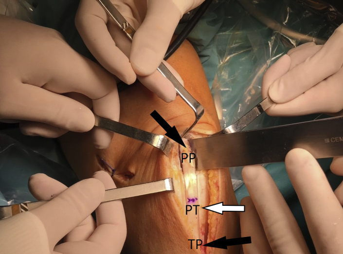
The osteotome is used to gently lever both bone plugs away from the remnant bone (right leg), producing the graft for final preparation. (PP, patellar plug; PT, patellar tendon; TP, tibial plug.)
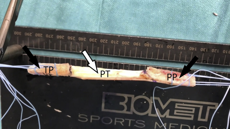
The aim is to harvest a tibial plug (TP) (10 mm × 25 mm), a patellar bone plug (PP) (10 mm × 30 mm), and an 11-mm-wide tendon graft (right leg). We leave at least 10 mm of patellar tendon (PT) medially and use a high-resistance suture (No. 2 FiberWire).

For bone-tendon-bone (BTB) graft placement, both sutures from the tibial tubercle bone plug are passed through the tibial and femoral tunnels with the passing suture (right leg).
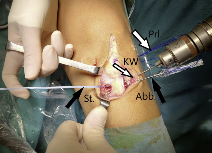
Two parallel transosseous tunnels are made with a 2-mm K-wire (KW) (left leg). The tunnels are made from the tibial groove (10 × 25 mm) to the place where the tibial tunnel begins (the sutures are located next to the interference screw), which should be lateral to the insertion of the patellar tendon and superior to the sartorius fascia. (Abb, Abbocath; Prl, Prolene suture; St, high-strength suture.)

A No. 1 Prolene suture (Prl) is passed through the 2 tunnels with the aid of 2 Abbocath devices; this will serve as a transport suture to pick up the high-strength suture (St) (No. 5 Ethibond) from the tibial plug and introduce it into the tibial groove (TG) (left leg). (TT, tibial tunnel.)

(A, B) We recommend a 1-cm separation between the 2 tunnels (left leg). (St, high-strength suture; TG, tibial groove; TT, tibial tunnel.)

(A, B) Both sutures are knotted (StK), and the knots are left in the tibial groove (TG), where they will not cause discomfort because they do not come into contact with the skin.
References
-
- Thompson S.M., Salmon L.J., Waller A., Linklater J., Roe J.P., Pinczewski L.A. Twenty-year outcome of a longitudinal prospective evaluation of isolated endoscopic anterior cruciate ligament reconstruction with patellar tendon or hamstring autograft. Am J Sports Med. 2016;44:3083–3094. - PubMed
-
- Tibor L., Chan P.H., Funahashi T.T., Wyatt R., Maletis G.B., Inacio M.C. Surgical technique trends in primary ACL reconstruction from 2007 to 2014. J Bone Joint Surg Am. 2016;98:1079–1089. - PubMed
-
- Fu F.H., Schulte K.R. Anterior cruciate ligament surgery 1996. State of the art? Clin Orthop Relat Res. 1996;(325):19–24. - PubMed
LinkOut - more resources
Full Text Sources
