

The value of narrow-band imaging bronchoscopy in diagnosing central lung cancer
- PMID: 36185220
- PMCID: PMC9524255
- DOI: 10.3389/fonc.2022.998770
The value of narrow-band imaging bronchoscopy in diagnosing central lung cancer
Abstract
Aims: This research aimed to study the value of narrow-band imaging(NBI) in the diagnosis of central lung cancer.
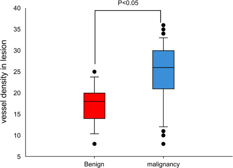
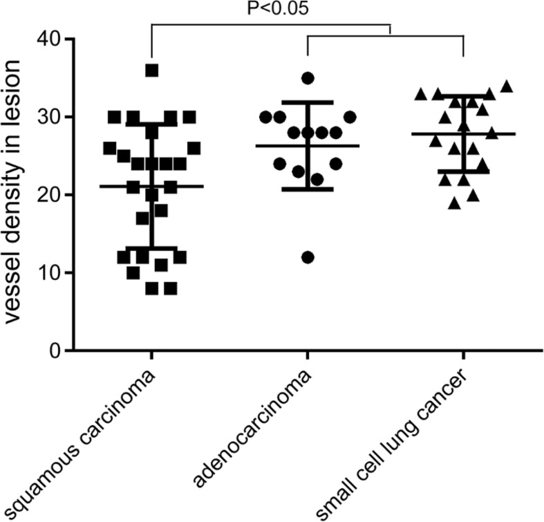
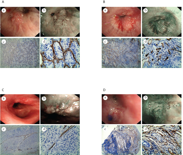
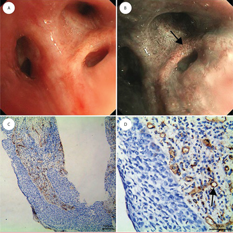
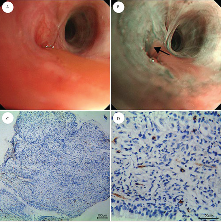
Materials and methods: This study included 916 patients with clinical suspected of central lung cancer or follow-up of patients after curative lung cancer surgery. All of the patients were examined by Olympus Evis Lucera electronic bronchoscope system, any sites that were abnormal when viewed by white-light bronchoscopy (WLB) or NBI were biopsied, four to six biopsies were taken at each site of the abnormal region visualized as lesions, we record the endoscopic features of NBI and compared with histopathology results, to evaluate the diagnostic value of NBI for central lung cancer and the relationship between vascular patterns of NBI and histological types of lung cancer, and try to establish a multinomial logistic regression model for predicting the histological types of lung cancer. The biopsy specimens were examined by CD34 antibody through immunohistochemistry (IHC) method, CD34 marked microvessel density(MVD), compared the number of microvessels between benign and malignant diseases and the number between different histological types of lung cancer, to verify the results of NBI.
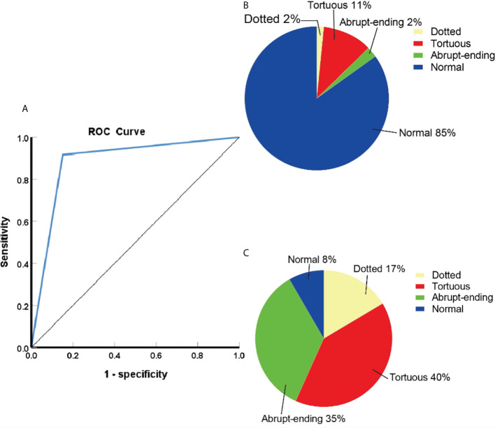
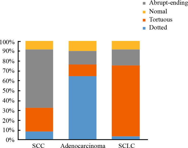
Results: NBI provided high sensitivity (91.7%), specificity (84.9%), positive predictive value (97.6%), negative predictive value (61.5%), and agreement rate (90.7%). The predominant vascular patterns in the well-defined histological types of lung cancer were dotted blood vessels (121 patients), tortuous blood vessels (248 patients), and abrupt-ending blood vessels (227 patients). Logistic regression analysis of the results showed that smoking status of the patient, combined with vascular patterns under NBI, and age partly affect the histological types of lung cancer.
Conclusions: NBI is highly accurate for the diagnosis of central lung cancer.
Keywords: CD34; central lung cancer; diagnosis; histological type; narrow-band imaging; vascular pattern.
Copyright © 2022 Zhu, Liu, Wu, Li, Gong, Shen, Ou and Li.
Conflict of interest statement
The authors declare that the research was conducted in the absence of any commercial or financial relationships that could be construed as a potential conflict of interest.
Figures
Similar articles
-
Relation between vascular patterns visualized by Narrow Band Imaging (NBI) videobronchoscopy and histological type of lung cancer.Med Oncol. 2013 Mar;30(1):374. doi: 10.1007/s12032-012-0374-x. Epub 2012 Dec 29. Med Oncol. 2013. PMID: 23275117 Clinical Trial.
-
Vascular patterns on narrow band imaging (NBI) video bronchoscopy of lung cancer patients and its relationship with histology: an analytical cross-sectional study.Adv Respir Med. 2021;89(1):30-36. doi: 10.5603/ARM.a2021.0014. Adv Respir Med. 2021. PMID: 33660246
-
[Combination of narrow-band imaging and autofluorescence imaging videobronchoscopy in the assessment of lung cancer].Zhongguo Fei Ai Za Zhi. 2013 Jun;16(6):299-302. doi: 10.3779/j.issn.1009-3419.2013.06.05. Zhongguo Fei Ai Za Zhi. 2013. PMID: 23769344 Free PMC article. Chinese.
-
[Narrow-band imaging bronchoscopy improves assessment of tumor extent and affects therapeutic strategy for central lung cancer].Zhonghua Yi Xue Za Zhi. 2014 Dec 2;94(44):3497-500. Zhonghua Yi Xue Za Zhi. 2014. PMID: 25622741 Chinese.
-
[Combination of narrow band imaging (NBI) and autofluorescence bronchoscopy (AFB) in the assessment of central lung cancer].Zhonghua Jie He He Hu Xi Za Zhi. 2014 Mar;37(3):184-7. Zhonghua Jie He He Hu Xi Za Zhi. 2014. PMID: 24809708 Chinese.
Cited by
-
The insufficiency of CT examination in early detection of central lung squamous cell carcinoma and squamous epithelial precancerous lesions.BMC Cancer. 2024 Mar 5;24(1):299. doi: 10.1186/s12885-024-12052-9. BMC Cancer. 2024. PMID: 38443800 Free PMC article.
-
A Deceptive Tracheal Mass Mimicking Asthma.J Brown Hosp Med. 2024 Jan 1;3(1):91419. doi: 10.56305/001c.91419. eCollection 2024. J Brown Hosp Med. 2024. PMID: 40027395 Free PMC article.
-
Feasibility of Multiparameter MRI-Guided Percutaneous Biopsy for Central Lung Lesions With Atelectasis.Korean J Radiol. 2025 May;26(5):498-507. doi: 10.3348/kjr.2024.0818. Korean J Radiol. 2025. PMID: 40307203 Free PMC article.
References
LinkOut - more resources
Full Text Sources