. 2022 Sep 15;25(10):105151.
doi: 10.1016/j.isci.2022.105151. eCollection 2022 Oct 21.

Cbl-b restrains priming of pathogenic Th17 cells via the inhibition of IL-6 production by macrophages

Affiliations

Cbl-b restrains priming of pathogenic Th17 cells via the inhibition of IL-6 production by macrophages

Qiuming Zeng et al. iScience. .

Abstract

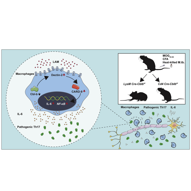
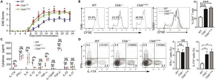
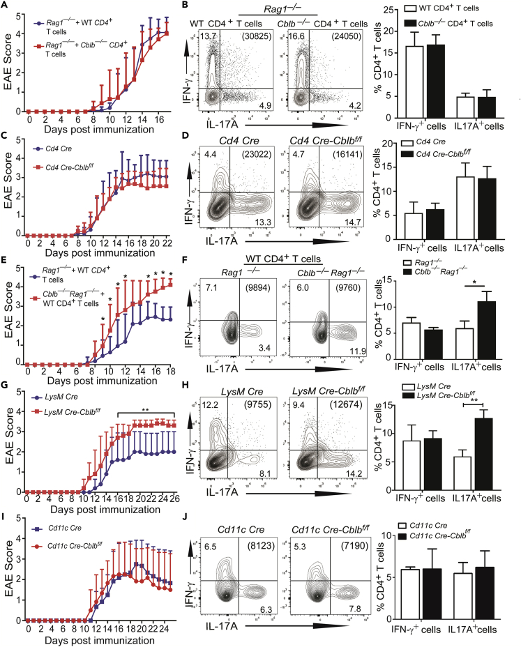
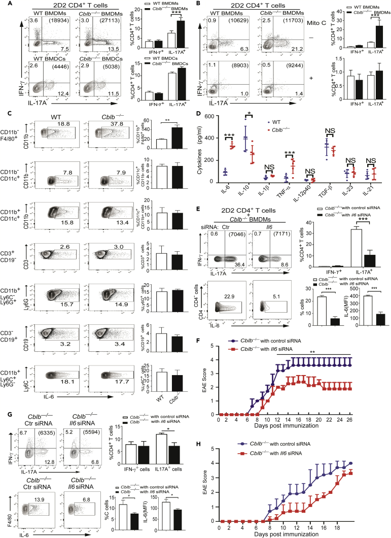
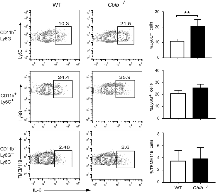
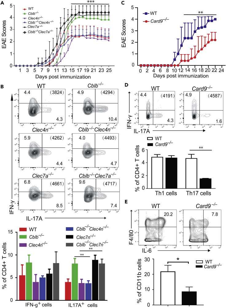
E3 ubiquitin ligase Cbl-b is involved in the maintenance of a balance between immunity and tolerance. Mice lacking Cbl-b are highly susceptible to experimental autoimmune encephalomyelitis (EAE), a Th17-mediated autoimmune disease. However, how Cbl-b regulates Th17 cell responses remains unclear. In this study, utilizing adoptive transfer and cell type-specific Cblb knockout strains, we show that Cbl-b expression in macrophages, but not T cells or dendritic cells (DCs), restrains the generation of pathogenic Th17 cells and the development of EAE. Cbl-b inhibits IL-6 production by macrophages that is induced by signaling from CARD9-dependent C-type lectin receptor (CLR) pathways, which directs T cells to generate pathogenic Th17 cells. Therefore, our data unveil a previously unappreciated function for Cbl-b in the regulation of pathogenic Th17 responses.

Keywords: Biological sciences; Immune response; Immunology.


Conflict of interest statement

The authors declare no competing financial interests.

Figures

None
Graphical abstract
Figure 1
Figure 1
Loss or inactivation of Cbl-b in mice leads to the development of severe clinical symptoms of EAE (A) Clinical scores of WT, Cblb−/−, and CblbC373A mice (n = 5/group) immunized with MOG35-55 in CFA. ∗p < 0.05; Mann-Whitney U test. (B and C) Ex vivo antigen-induced T cell proliferation and cytokine production of WT, Cblb−/−, and CblbC373A mice (n = 3/group for B, n = 5/group for C) immunized with MOG35-55 in CFA as described in A, determined by CFSE labeling and ELISA. ∗∗p < 0.01, ∗∗∗p < 0.001; Student t test. (D) Flow cytometric analysis of Th1/Th17 responses in dLNs of WT, Cblb−/−, and CblbC373A mice (n = 3/group) on day 8 after immunization with MOG35-55 in CFA. ∗∗p < 0.01; Student t test. Bracket: the total number of gated CD4+ T cells. Data are one representative of three independent experiments.
Figure 2
Figure 2
Cbl-b deficiency in myeloid cells is responsible for heightened EAE and Th17 responses (A) Clinical scores of Rag1−/− mice receiving CD4+ T cells from WT (n = 8) and Cblb−/− (n = 7) mice, immunized with MOG35-55 in CFA. (B) Flow cytometric analysis of Th1/Th17 responses in dLNs of Rag1−/−mice receiving CD4+ T cells from WT and Cblb−/− mice (n = 3/group) on day 8 after immunization with MOG35-55 in CFA. (C) Clinical scores of Cd4 Cre (n = 10) and Cd4 Cre-Cblbf/f (n = 9) mice, immunized with MOG35-55 in CFA. (D) Flow cytometric analysis of Th1/Th17 responses in dLNs of Cd4 Cre and Cd4 Cre-Cblbf/f mice (n = 3/group) on day 8 after immunization with MOG35-55 in CFA. (E) Clinical scores of Rag1−/− (n = 11) and Cblb−/−Rag1−/− (n = 10) mice receiving CD4+ T cells from WT mice, immunized with MOG35-55 in CFA. ∗p < 0.05; Mann-Whitney U test. (F) Flow cytometric analysis of Th1/Th17 responses in dLNs of Rag1−/− and Cblb−/−Rag1−/− mice (n = 4/group) receiving CD4+ T cells from WT mice on day 8 after immunization with MOG35-55 in CFA. ∗p < 0.05; Student t test. (G) Clinical scores of LysM Cre and LysM Cre-Cblbf/f mice (n = 8/group), immunized with MOG35-55 in CFA. ∗∗p < 0.01; Mann-Whitney U test. (H) Flow cytometric analysis of Th1/Th17 responses in dLNs of LysM Cre and LysM Cre-Cblbf/f mice (n = 3/group) on day 8 after immunization with MOG35-55 in CFA. ∗∗p < 0.01; Student t test. (I) Clinical scores of Cd11c Cre and Cd11c Cre-Cblbf/f mice (n = 6/group), immunized with MOG35-55 in CFA. (J) Flow cytometric analysis of Th1/Th17 responses in dLNs of Cd11c Cre and Cd11c Cre-Cblbf/f mice (n = 3/group) on day 8 after immunization with MOG35-55 in CFA. Bracket: the total number of gated CD4+ T cells. Data are representative of two independent experiments.
Figure 3
Figure 3
Hyper-production of IL-6 by Cblb−/− macrophages promotes a pathogenic Th17 response (A) Th1/Th17 cell differentiation in co-cultures of 2D2 T cells with BMDMs or BMDCs from WT and Cblb−/− mice (n = 3/group) in the presence of heat-killed M.tb and MOG35-55. ∗∗∗p < 0.001; Student t test. (B) Th1/Th17 cell differentiation in co-cultures of 2D2 T cells with BMDMs from WT and Cblb−/− mice (n = 3/group) in the presence of heat-killed M.tb and MOG35-55 with or without pretreatment with mitomycin C. ∗∗∗p < 0.001; Student t test. (C) Flow cytometric analysis of IL-6-producing cells of dLNs of WT and Cblb−/− mice (n = 3/group) on day 8 after immunization with MOG35-55 in CFA, and stimulated with PMA/ionomycin. The gating strategy and representative plots see Figure S5. ∗∗p < 0.01; Student t test. (D) ELISA of pro-inflammatory cytokines (TNF-α, IL-6, IL-10, IL-12p40, IL-1β, TGF-β, IL-23, and IL-21) by BMDMs from WT and Cblb−/− mice (n = 5/group) stimulated with heat-killed M.tb. ∗p < 0.05, ∗∗∗p < 0.001; Student t test. (E) Th1/Th17 cell differentiation in co-cultures of 2D2 CD4+ T cells with BMDMs from Cblb−/− mice that had been treated with Il6 or control siRNA. IL-6-producing macrophages of Cblb−/− mice treated with Il6 (n = 4) or control siRNA (n = 4) were also determined. The mean fluorescent intensity (MFI) determined by flow cytometry indicates the expression level of IL-6 in CD4 macrophages of Cblb−/− mice treated with Il6 or control siRNA. ∗∗∗p < 0.001; Student t test. (F) Clinical scores of Cblb−/− mice immunized with MOG35-55 in CFA in the treatment with or without Il6 siRNA (n = 5) or control siRNA (n = 5) on day 1, 3, and 7 via tail vein injection. ∗∗p < 0.01; Mann-Whitney U test. (G) Flow cytometric analysis of Th1/Th17 responses and IL-6-producing cells in dLN cells from Cblb−/− mice treated with Il6 (n = 3) or control siRNA (n = 3) via tail vein injection as in F on day 8 after immunization with MOG35-55 in CFA. MFI determined by flow cytometry indicates the expression level of IL-6 in F4/80+ cells from dLNs of Cblb−/− mice treated with Il6 (n = 3) or control siRNA (n = 3). ∗p < 0.05; Student t test. (H) Clinical scores of Cblb−/− mice immunized with MOG35-55 in CFA in the treatment with or without Il6 siRNA (n = 5) or control siRNA (n = 5) on day 8, 11, and 15 via tail vein injection. Bracket: the total number of gated CD4+ T cells. Data are representative of three independent experiments (A, B, E, F, and G) and representative of two independent experiments (C-D).
Figure 4
Figure 4
CNS macrophages, but not neutrophils and microglia, from Cblb–/– mice with EAE produce more IL-6 Flow cytometric analysis of IL-6-producing macrophages (CD11b+Ly6C+Ly6G), neutrophils(CD11b+Ly6G+Ly6C+), and microglia (CD11b+Ly6GLy6CTMEM119+) cells of CNS of WT and Cblb−/− mice (n = 4/group) on day 17 after immunization with MOG35-55 in CFA, and stimulated with PMA/ionomycin. ∗∗p < 0.01; Student t test. Data are representative of two independent experiments.
Figure 5
Figure 5
CARD9-mediated Dectin receptor signaling contributes the clinical symptoms of Cblb−/− mice with EAE and pathogenic Th17 response (A) Clinical scores of WT, Cblb−/−, Clec4n−/−, Clec7a−/−, Cblb−/−Clec4n−/−, and Cblb−/−Clec7a−/− mice (n = 5/group) immunized with immunized with MOG35-55 in CFA. ∗∗∗p < 0.001, Mann-Whitney U test. (B) Flow cytometric analysis of Th1/Th17 responses in dLNs of WT, Cblb−/−, Clec4n−/−, Clec7a−/−, Cblb−/−Clec7a−/−, and Cblb−/−Clec4n−/− mice (n = 4/group) on day 8 after immunization with MOG35-55 in CFA. ∗∗p < 0.01, Student t test. (C) Clinical scores of WT and Card9−/− mice (n = 7/group) immunized with MOG35-55 in CFA. ∗∗p < 0.01, Mann-Whitney U test. (D) Flow cytometric analysis of Th1/Th17 responses in dLNs WT and Card9−/− mice (n = 5/group) on day 8 after immunization with MOG35-55 in CFA. ∗∗p < 0.01, Student’s t test. (E) Flow cytometric analysis of Th1/Th17 responses in dLNs WT and Card9−/− mice (n = 3/group) on day 8 after immunization with MOG35-55 in CFA. ∗p < 0.05, Student’s t test. Data are three independent experiments for A and B, and two independent experiments for C, D, and E.

Similar articles

Cited by

References

    1. Alonso R., Fernandez-Fernandez A.M., Pisa D., Carrasco L. Multiple sclerosis and mixed microbial infections. Direct identification of fungi and bacteria in nervous tissue. Neurobiol. Dis. 2018;117:42–61. - PubMed
    1. Aqel S.I., Yang X., Kraus E.E., Song J., Farinas M.F., Zhao E.Y., Pei W., Lovett-Racke A.E., Racke M.K., Li C., Yang Y. A STAT3 inhibitor ameliorates CNS autoimmunity by restoring Teff:Treg balance. JCI Insight. 2021;6 - PMC - PubMed
    1. Bachmaier K., Krawczyk C., Kozieradzki I., Kong Y.Y., Sasaki T., Oliveira-dos-Santos A., Mariathasan S., Bouchard D., Wakeham A., Itie A., et al. Negative regulation of lymphocyte activation and autoimmunity by the molecular adaptor Cbl-b. Nature. 2000;403:211–216. - PubMed
    1. Bergholdt R., Taxvig C., Eising S., Nerup J., Pociot F. CBLB variants in type 1 diabetes and their genetic interaction with CTLA4. J. Leukoc. Biol. 2005;77:579–585. - PubMed
    1. Bettelli E., Pagany M., Weiner H.L., Linington C., Sobel R.A., Kuchroo V.K. Myelin oligodendrocyte glycoprotein-specific T cell receptor transgenic mice develop spontaneous autoimmune optic neuritis. J. Exp. Med. 2003;197:1073–1081. - PMC - PubMed

LinkOut - more resources