. 2022 Sep 9;25(10):105106.
doi: 10.1016/j.isci.2022.105106. eCollection 2022 Oct 21.

Underwater instant adhesion mechanism of self-assembled amphiphilic hemostatic granular hydrogel from Andrias davidianus skin secretion

Affiliations

Underwater instant adhesion mechanism of self-assembled amphiphilic hemostatic granular hydrogel from Andrias davidianus skin secretion

Yuqing Liu et al. iScience. .

Abstract

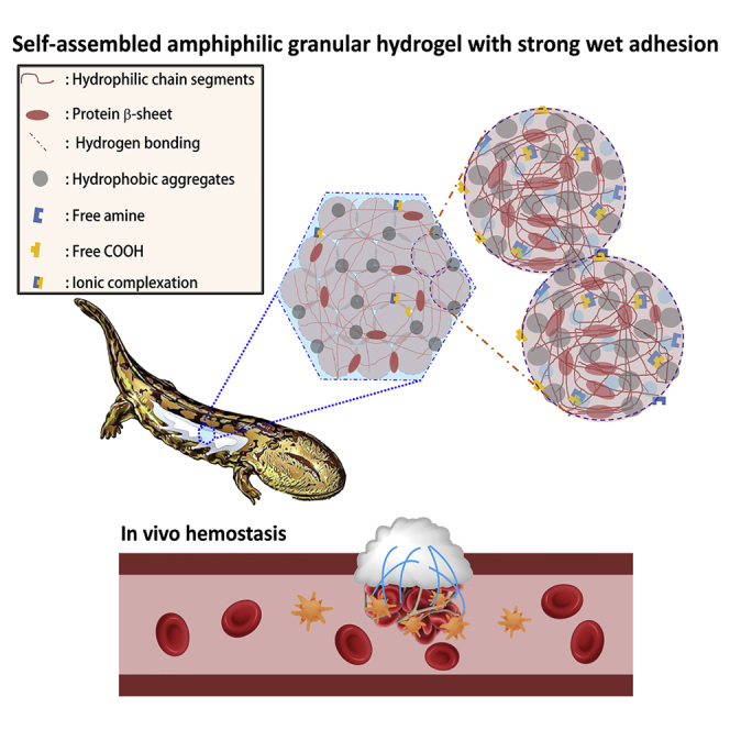
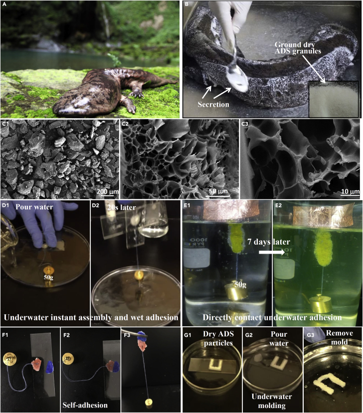
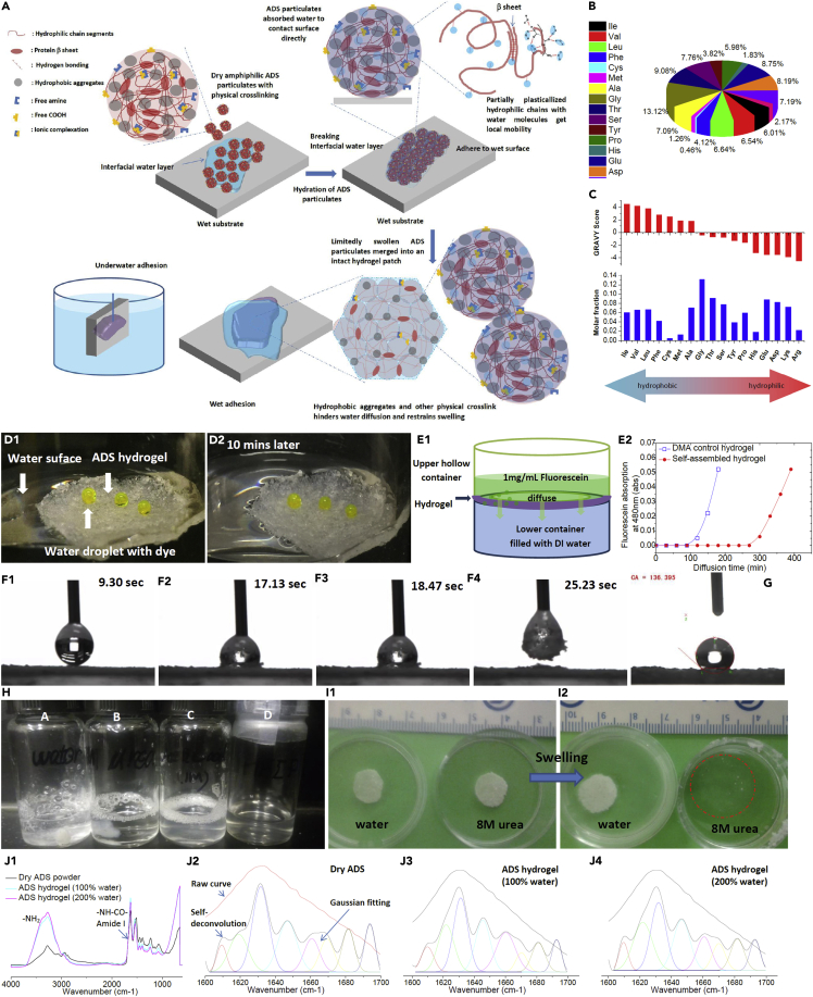
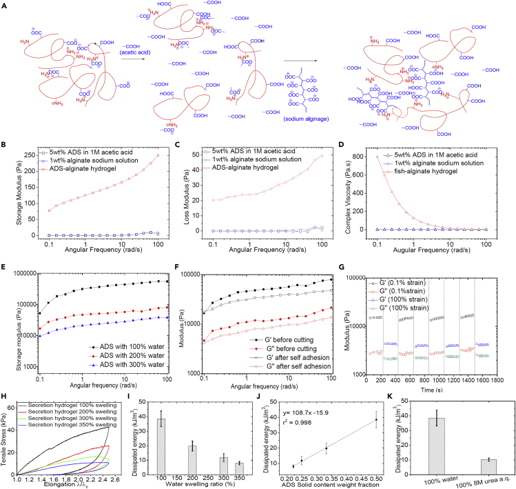
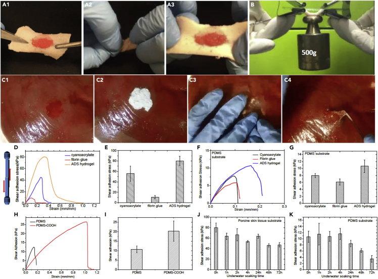
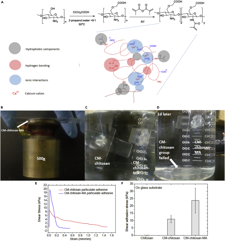
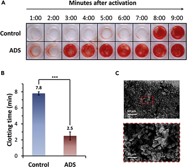
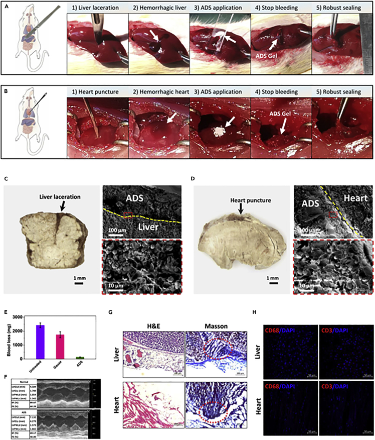
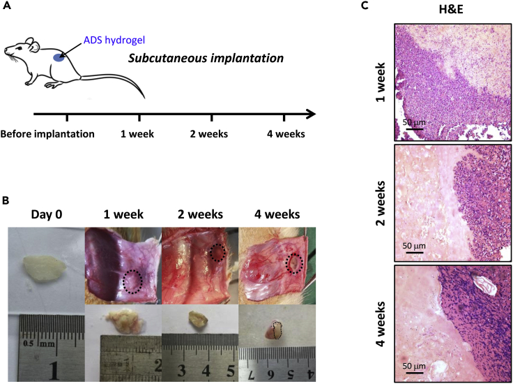
The widespread use of biological tissue adhesives for tissue repair is limited by their weak adhesion in a wet environment. Herein, we report the wet adhesion mechanism of a dry granular natural bioadhesive from Andrias davidianus skin secretion (ADS). Once contacting water, ADS granules self-assemble to form a hydrophobic hydrogel strongly bonding to wet substrates in seconds. ADS showed higher shear adhesion than current commercial tissue adhesives and an impressive 72-h underwater adhesion strength of ∼47kPa on porcine skin tissue. The assembled hydrogel in water maintained a dissipation energy of ∼8 kJ/m3, comparable to the work density of muscle, exhibiting its robustness. Unlike catechol adhesion mechanism, ADS wet adhesion mechanism is attributed to water absorption by granules, and the unique equilibrium of protein hydrophobicity, hydrogen bonding, and ionic complexation. The in vivo adhesion study demonstrated its excellent wet adhesion and hemostasis performance in a rat hepatic and cardiac hemorrhage model.

Keywords: Biomaterials; Materials chemistry; Materials science.


Conflict of interest statement

The authors declare no competing interests.

Figures

None
Graphical abstract
Figure 1
Figure 1
Underwater contact adhesion behavior of instantly self-assembled granular hydrogels from dry ADS particulates (A) Digital photograph of an adult Andrias Davidianus. (B) Digital photograph of the skin secretion collection (the inset shows ADS powders). (C) SEM images of dry ADS granules (c1, scale bar: 200 μm) and self-assembled ADS granular hydrogel internal cross-section at different magnifications (c2 and c3, scale bar: 50 μm and 10 μm respectively). (D) Instant self-assembly of ADS particulates to form an adhesive patch in water (E) 50 g weight handing from the hydrogel was soaked in water for up to 7 days (the color was from the fluorescein dye). (F) Self-adhesion behavior of ADS hydrogel and the repaired patch could afford a weight of 20 g. (G) Molding ADS into a U-shape hydrogel via self-assembly and self-adhesion.
Figure 2
Figure 2
Wet adhesion mechanism of ADS (A) Schematic illustration of dry ADS particulates’ self-assembly and adhesion mechanism in water. (B) The amino acid composition analysis of ADS proteins. (C) The GRAVY score evaluation of ADS proteins. (D) The hydrophobic behavior of self-assembled hydrogels floated on water. (E) The diffusion behavior of fluorescein dye a.q. solution (1 mg/mL) passing through an ADS hydrogel patch or a control hydrogel (dimethylacrylamide, DMA) (thickness= 1mm). (F) Hydration and hydrophobic self-assembly of ADS particles on the air-water interface of a water droplet. (G) The water contact angle of a water droplet on ADS hydrogel surface. (H) The solubility of ADS hydrogel (100% water-swollen, 20mg) in different solvents (1 mL). (A) water, (B) 8M urea a.q. solution, (C) 1M acetic acid solution, and (D) HFIP. (I) The swelling behaviors of the ADS hydrogel (100% original swollen ratio) in water or 8M urea a.q. solution respectively. (J1) The FTIR-ATR spectra of dry ADS powder and ADS hydrogel with different hydration levels (100% or 200%). The FTIR-ATR raw curves, self-deconvoluted curves, and corresponding Gaussian fittings in the amide I region of dry ADS powder (J2), 100% swollen ADS hydrogel (J3), or 200% swollen ADS hydrogel (J4).
Figure 3
Figure 3
Physical crosslinking in ADS hydrogel (A) Schematic illustration of ionic complexation contribution in ADS hydrogel. (B–D) The rehological profiles of 5wt% ADS in 1M acetic acid solution, 1wt% alginate aqueous solution, and the mixture of ADS-acetic acid/alginate solution in oscillation frequency sweeping test, including storage modulus vs. angular frequency (B), loss modulus vs. angular frequency (C) and complex viscosity vs. angular frequency (D). (E) Frequency sweep rheological profiles of ADS hydrogel with different swelling ratios (100, 200, and 300%). (F) Frequency sweep test of ADS hydrogel before cutting and after self-adhesion. (G) The dynamic strain amplitude alternating cyclic test of ADS hydrogel at the shearing rate of 1 rad/s, the strains were shifted between 0.1% or 100% for 8 steps (4 cycles), and the period per step was 200 s. (H)The tensile stress-strain cycle curves of ADS hydrogels with different swelling ratios. (I) The dissipated energy of ADS hydrogels with different swelling ratios (n = 3). (J) The linearity of dissipated energy vs. ADS solid content (n = 3). (K) Comparison of dissipated energy between ADS hydrogels swollen with 100% water or 8M urea a.q. solution (n = 3).
Figure 4
Figure 4
The adhesion performance of ADS hydrogel (A) ADS hydrogel adhered to porcine skin tissue twisted and restored. (B) ADS hydrogel formed on porcine skin tissue surface bearing a weight of 500 g. (C) ADS hydrogel in-situ formed and adhered to the porcine liver surface via self-assembly. (D and E) Lap shear adhesion stress of ADS hydrogel, cyanoacrylate, and fibrin glue on porcine skin tissue substrate (n = 3 ). (F and G) Lap shear adhesion stress of ADS hydrogel, cyanoacrylate adhesive, and fibrin glue on PDMS substrate (n = 3). (H and I) Lap shear adhesion of ADS hydrogel on regular PDMS or carboxylic acids decorated PDMS (PDMS-COOH) surface (n = 3). (J and K) Underwater contact adhesion of ADS assembled hydrogel on porcine skin tissue and PDMS substrate surface (n = 3).
Figure 5
Figure 5
Synthetic model polymer methacrylated carboxymethyl chitosan (CM-chitosan-MA) granular adhesive to mimic the wet adhesion behavior of ADS hydrogel (A) The designed model polymer, CM-chitosan-MA, based granular hydrogel adhesive. (B) CM-chitosan-MA particulates with 100% water self-assembled to a hydrogel patch to hold a weight of 500 g on a glass substrate. (C and D) Underwater adhesion performance of CM-chitosan-MA particulates and CM-chitosan particulates (control group). (E and F) Lap shear adhesion of CM-chitosan-MA and CM-chitosan hydrogels on glass substrates. Note: with water, chitosan particles could not assemble and could not adhere to substrates (n = 3).
Figure 6
Figure 6
Characterization of the procoagulant effect of ADS on the whole blood (A) Qualitative analysis of blood clotting with or without ADS powder treatment in 96-well plates. (B) Quantification of clotting time of whole blood in contact with ADS powder compared to control group. Data are means ± SD (n = 3). p value determined by Student’s ttest (∗∗∗p < 0.001). (C) SEM analysis of the interaction of ADS with red blood cells (RBCs, scale bar: 50 μm and 10 μm respectively).
Figure 7
Figure 7
Hemostatic performance of ADS hydrogel in rat’s hepatic and cardiac hemorrhage model (A and B) The rapid hemostasis and sealing process of ADS hydrogel for rat liver laceration (A) and heart puncture (B); (C and D) Photographic (left) and SEM (right) images of freeze-fractured ADS-tissue interface for injured liver (C) and heart (D) tissues. The interfaces were marked with a dotted yellow line (scale bar: 1mm (left), 100 μm (right top), and 10 μm (right bottom) respectively). (E) Quantification of blood loss for lacerated liver treated with ADS hydrogel, gauze and control (untreated) (n = 3). (F) Echocardiography measurements of normal and ADS-treated groups after 4 weeks. (G) Histology images of ADS hydrogel and the around injured tissue stained with H&E and Masson’s trichrome after 4 weeks of treatment (scale bar: 200 μm). (H) Immunostaining images (DAPI, CD3 and CD68 marker) of injured tissue treated with ADS hydrogel after 4 weeks (scale bar: 50 μm).
Figure 8
Figure 8
In vivo biocompatibility and biodegradability evaluation (A) Schematic of ADS implantation in the rat dorsal subcutaneous pocket. (B) Macroscopic appearance of ADS hydrogel pre-implantation and post-implantation excised at 1, 2 and 4 weeks. (C) Histological images of ADS hydrogel implants and surrounding tissues after 1, 2 and 4 weeks after implantation (scale bar: 50 μm).

Similar articles

Cited by

References

    1. Annabi N., Zhang Y.N., Assmann A., Sani E.S., Cheng G., Lassaletta A.D., Vegh A., Dehghani B., Ruiz-Esparza G.U., Wang X., et al. Engineering a highly elastic human protein-based sealant for surgical applications. Sci. Transl. Med. 2017;9:eaai7466. - PMC - PubMed
    1. Avery R.K., Albadawi H., Akbari M., Zhang Y.S., Duggan M.J., Sahani D.V., Olsen B.D., Khademhosseini A., Oklu R. An injectable shear-thinning biomaterial for endovascular embolization. Sci. Transl. Med. 2016;8:365ra156. doi: 10.1126/scitranslmed.aah5533. - DOI - PubMed
    1. Baik S., Kim D.W., Park Y., Lee T.-J., Ho Bhang S., Pang C. A wet-tolerant adhesive patch inspired by protuberances in suction cups of octopi. Nature. 2017;546:396–400. doi: 10.1038/nature22382. - DOI - PubMed
    1. Barton A.F.M. Second edition. CRC Press; 2017. CRC Handbook of Solubility Parameters and Other Cohesion Parameters.
    1. Bouten P.J., Zonjee M., Bender J., Yauw S.T., van Goor H., van Hest J.C., Hoogenboom R. The chemistry of tissue adhesive materials. Prog. Polym. Sci. 2014;39:1375–1405. doi: 10.1016/j.progpolymsci.2014.02.001. - DOI

LinkOut - more resources