Review
. 2022 Sep 14:10:981061.
doi: 10.3389/fbioe.2022.981061. eCollection 2022.

Perinatal derivatives: How to best validate their immunomodulatory functions

Affiliations
Review

Perinatal derivatives: How to best validate their immunomodulatory functions

Andrea Papait et al. Front Bioeng Biotechnol. .

Abstract

Perinatal tissues, mainly the placenta and umbilical cord, contain a variety of different somatic stem and progenitor cell types, including those of the hematopoietic system, multipotent mesenchymal stromal cells (MSCs), epithelial cells and amnion epithelial cells. Several of these perinatal derivatives (PnDs), as well as their secreted products, have been reported to exert immunomodulatory therapeutic and regenerative functions in a variety of pre-clinical disease models. Following experience with MSCs and their extracellular vesicle (EV) products, successful clinical translation of PnDs will require robust functional assays that are predictive for the relevant therapeutic potency. Using the examples of T cell and monocyte/macrophage assays, we here discuss several assay relevant parameters for assessing the immunomodulatory activities of PnDs. Furthermore, we highlight the need to correlate the in vitro assay results with preclinical or clinical outcomes in order to ensure valid predictions about the in vivo potency of therapeutic PnD cells/products in individual disease settings.

Keywords: exosomes; extracellular vesicles; functional assays; immunomodulation; mechanisms of action; mesenchymal stromal cells; microvesicles; perinatal derivatives.


Conflict of interest statement

The authors declare that the research was conducted in the absence of any commercial or financial relationships that could be construed as a potential conflict of interest.

Figures

FIGURE 1
FIGURE 1
Analysis of T-lymphocyte function. (A) T lymphocytes can be activated using various stimuli including monoclonal antibodies (stimulation with anti-CD3 anti-CD28), and mitogens, such as PMA frequently used in combination with ionomycin. Other modes of stimulation include the use of lipopolysaccharides that triggers TLR4 activation (thus mimicking the bacterial stimulus), and causing the release of PHA that induces the recruitment of TCRs consequently activating T lymphocytes. Finally, T lymphocytes can also be stimulated by ConA, which causes the release of intracellular Ca2+ that triggers the calcium cascade, and by mixed lymphocyte reactions which are based on the allogeneic response determined by HLA mismatching between two different donors. (B) Depending on the considered mechanism of action, different readout methods are used. Flow cytometry can perform in-depth immune-phenotype analyses. (C) Various markers can be used to analyze T cell activity in given assays, some being selectively expressed at specific timepoints following T cell activation. (D) The functional polarization of T lymphocytes can be triggered with different combinations of cytokines towards different CD4 Th subsets or towards different CD8 memory T cell subsets. Cytokine analyses provide important information about resulting T cell functions. (Created with BioRender.com).
FIGURE 2
FIGURE 2
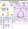
Impact of PnD on monocyte differentiation towards antigen presenting cells. (A) Perinatal Derivatives (PnD) and their secreted factors impact monocyte differentiation towards antigen presenting cells fostering the acquisition of phenotype and functional features typical of M2 macrophages. (B) Depending on the factors present in the microenvironment monocytes can be discriminated into different subsets of M2 macrophages (M2a, M2b, M2c and M2d) each of them being characterized by peculiar functions. (C) Summary table for the markers specific for macrophage and DC subsets. (D) Summary table for the cytokines released by the different macrophage/DC subsets. (Created with BioRender.com).

References

    1. Abraham R. T., Weiss A. (2004). Jurkat T cells and development of the T-cell receptor signalling paradigm. Nat. Rev. Immunol. 4 (4), 301–8. 10.1038/nri1330 - DOI - PubMed
    1. Arneth B. M. (2010). Measurement of T cell activation after 16-hr in vitro stimulation with concanavalin A. Curr. Protoc. Cytom. 6, 1–10. 10.1002/0471142956.cy0628s51 - DOI - PubMed
    1. Bai F., Zhang P., Fu Y., Chen H., Zhang M., Huang Q., et al. (2020). Targeting ANXA1 abrogates Treg-mediated immune suppression in triple-negative breast cancer. J. Immunother. Cancer 8 (1), e000169. 10.1136/jitc-2019-000169 - DOI - PMC - PubMed
    1. Bailo M., Soncini M., Vertua E., Signoroni P. B., Sanzone S., Lombardi G., et al. (2004). Engraftment potential of human amnion and chorion cells derived from term placenta. Transplantation 78 (10), 1439–1448. 10.1097/01.tp.0000144606.84234.49 - DOI - PubMed
    1. Balza E., Piccioli P., Carta S., Lavieri R., Gattorno M., Semino C., et al. (2016). Proton pump inhibitors protect mice from acute systemic inflammation and induce long-term cross-tolerance. Cell. Death Dis. 7 (7), e2304. 10.1038/cddis.2016.218 - DOI - PMC - PubMed

LinkOut - more resources