

Retinal organoids and microfluidic chip-based approaches to explore the retinitis pigmentosa with USH2A mutations
- PMID: 36185441
- PMCID: PMC9524156
- DOI: 10.3389/fbioe.2022.939774
Retinal organoids and microfluidic chip-based approaches to explore the retinitis pigmentosa with USH2A mutations
Abstract
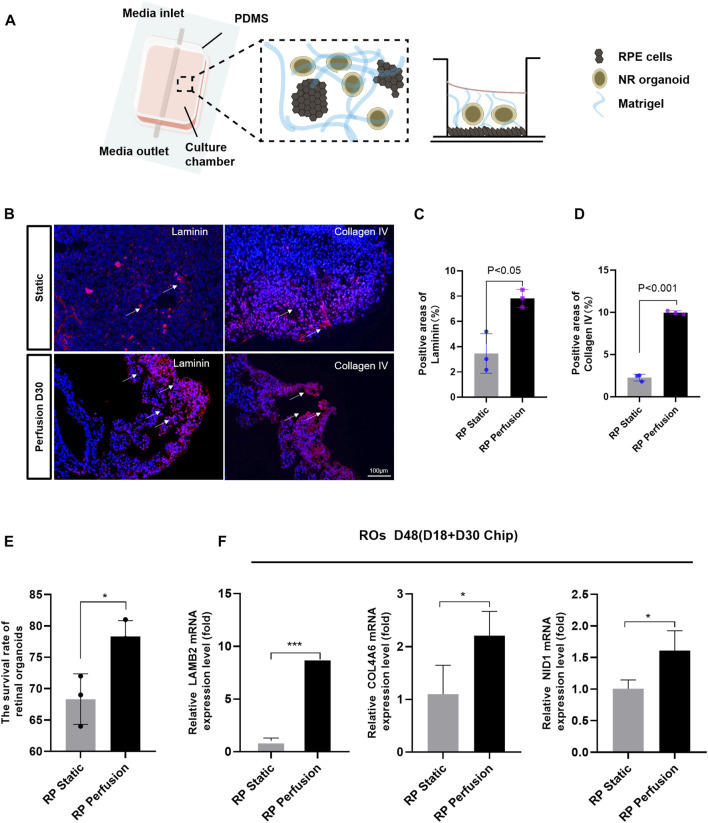
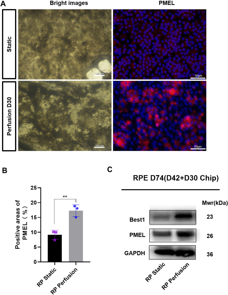
Retinitis pigmentosa (RP) is a leading cause of vision impairment and blindness worldwide, with limited medical treatment options. USH2A mutations are one of the most common causes of non-syndromic RP. In this study, we developed retinal organoids (ROs) and retinal pigment epithelium (RPE) cells from induced pluripotent stem cells (iPSCs) of RP patient to establish a sustainable in vitro RP disease model. RT-qPCR, western blot, and immunofluorescent staining assessments showed that USH2A mutations induced apoptosis of iPSCs and ROs, and deficiency of the extracellular matrix (ECM) components. Transcriptomics and proteomics findings suggested that abnormal ECM-receptor interactions could result in apoptosis of ROs with USH2A mutations via the PI3K-Akt pathway. To optimize the culture conditions of ROs, we fabricated a microfluidic chip to co-culture the ROs with RPE cells. Our results showed that this perfusion system could efficiently improve the survival rate of ROs. Further, ECM components such as laminin and collagen IV of ROs in the RP group were upregulated compared with those maintained in static culture. These findings illustrate the potential of microfluidic chip combined with ROs technology in disease modelling for RP.
Keywords: USH2A; induced pluripoten stem cells; microfludic chip; retinal organoids; retinitis pigmentosa.
Copyright © 2022 Su, Liang, Zhang, Wang, Chen, Su, Cao, Yu, Deng, Chan, Tang, Guo and Chen.
Conflict of interest statement
The authors declare that the research was conducted in the absence of any commercial or financial relationships that could be construed as a potential conflict of interest.
Figures


Similar articles
-
Modeling Retinitis Pigmentosa: Retinal Organoids Generated From the iPSCs of a Patient With the USH2A Mutation Show Early Developmental Abnormalities.Front Cell Neurosci. 2019 Aug 7;13:361. doi: 10.3389/fncel.2019.00361. eCollection 2019. Front Cell Neurosci. 2019. PMID: 31481876 Free PMC article.
-
USH2A variants causing retinitis pigmentosa or Usher syndrome provoke differential retinal phenotypes in disease-specific organoids.HGG Adv. 2023 Aug 7;4(4):100229. doi: 10.1016/j.xhgg.2023.100229. eCollection 2023 Oct 12. HGG Adv. 2023. PMID: 37654703 Free PMC article.
-
Visual Prognosis in USH2A-Associated Retinitis Pigmentosa Is Worse for Patients with Usher Syndrome Type IIa Than for Those with Nonsyndromic Retinitis Pigmentosa.Ophthalmology. 2016 May;123(5):1151-60. doi: 10.1016/j.ophtha.2016.01.021. Epub 2016 Feb 27. Ophthalmology. 2016. PMID: 26927203
-
Genetic dissection of non-syndromic retinitis pigmentosa.Indian J Ophthalmol. 2022 Jul;70(7):2355-2385. doi: 10.4103/ijo.IJO_46_22. Indian J Ophthalmol. 2022. PMID: 35791117 Free PMC article. Review.
-
Non-syndromic retinitis pigmentosa.Prog Retin Eye Res. 2018 Sep;66:157-186. doi: 10.1016/j.preteyeres.2018.03.005. Epub 2018 Mar 27. Prog Retin Eye Res. 2018. PMID: 29597005 Review.
Cited by
-
"Armed in-vitro retina"-generating microglial retinal organoids, where are we now?Front Cell Dev Biol. 2025 May 30;13:1574283. doi: 10.3389/fcell.2025.1574283. eCollection 2025. Front Cell Dev Biol. 2025. PMID: 40519267 Free PMC article. Review.
-
Current approaches for Usher syndrome disease models and developing therapies.Front Cell Dev Biol. 2025 Jun 20;13:1547523. doi: 10.3389/fcell.2025.1547523. eCollection 2025. Front Cell Dev Biol. 2025. PMID: 40620763 Free PMC article. Review.
-
Retinal Organoids from Induced Pluripotent Stem Cells of Patients with Inherited Retinal Diseases: A Systematic Review.Stem Cell Rev Rep. 2025 Jan;21(1):167-197. doi: 10.1007/s12015-024-10802-7. Epub 2024 Oct 18. Stem Cell Rev Rep. 2025. PMID: 39422807 Free PMC article.
-
Advanced Cellular Models for Rare Disease Study: Exploring Neural, Muscle and Skeletal Organoids.Int J Mol Sci. 2024 Jan 13;25(2):1014. doi: 10.3390/ijms25021014. Int J Mol Sci. 2024. PMID: 38256087 Free PMC article. Review.
-
Exploring organoid and assembloid technologies: a focus on retina and brain.Expert Rev Mol Med. 2025 Mar 27;27:e14. doi: 10.1017/erm.2025.9. Expert Rev Mol Med. 2025. PMID: 40145178 Free PMC article. Review.
References
LinkOut - more resources
Full Text Sources