Case Reports
. 2022 Aug 5;7(3):240-246.
doi: 10.14744/bej.2022.94546. eCollection 2022.

Unilateral Endogenous Bacterial Endophthalmitis Post-Coronavirus Disease-19 in an Healthy Asian Indian Male

Affiliations
Case Reports

Unilateral Endogenous Bacterial Endophthalmitis Post-Coronavirus Disease-19 in an Healthy Asian Indian Male

Srinivasan Sanjay et al. Beyoglu Eye J. .

Abstract

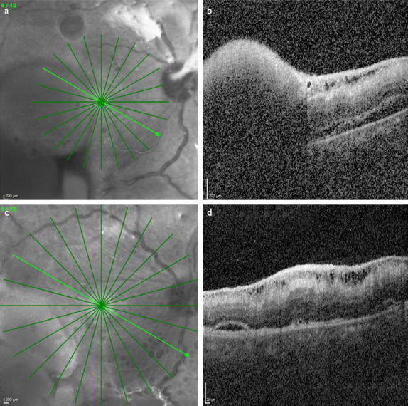
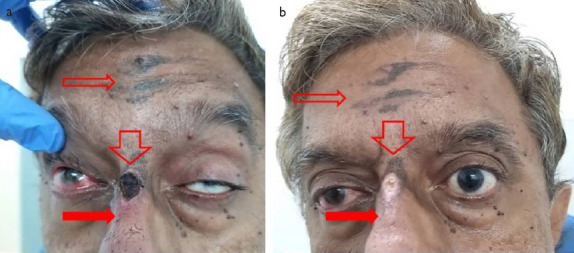
Coronavirus disease 2019 (COVID-19) is associated with ocular involvement either during or after the infection. These include conjunctivitis, conjunctival hyperemia, chemosis, epiphora, reactivation of anterior uveitis, or presenting as anterior sclero-uveitis, cotton wool spots, retinal hemorrhages, retinal artery/vein occlusion, ophthalmic artery occlusion, panuveitis, papillophlebitis, central serous retinopathy, presumed fungal endophthalmitis, and multifocal chorioretinitis. A 47-year-old Asian Indian male was diagnosed with COVID-19 and had no other systemic history of note at the time of admission. Three weeks later, he developed sudden loss of vision in the right eye (OD). Visual acuity in OD was perception of light. OD had features of endophthalmitis. OD underwent pars plana vitrectomy with intravitreal antibiotics. Anterior chamber tap for fungal culture and polymerase chain reaction for panfungal genome was negative. Culture of ocular specimens did not reveal bacterial growth. Vitreous sample showed few Gram-positive cocci in singles and pairs with no evidence of fungal elements. Polymerase chain reaction for eubacterial genome was positive. He was treated with topical and systemic antibiotics and steroids. Final follow-up 6 weeks later, OD had a best-corrected visual acuity which was 20/200 with a quiet anterior chamber, cataract, with a macular traction and reduced sub retinal exudates and fluid. Post-COVID-19 sequelae causing sight-threatening manifestations as illustrated by this case report needs early recognition and prompt treatment to achieve a favorable visual outcome.

Keywords: Coronavirus disease-19; endogenous endophthalmitis; pars plana vitrectomy; polymerase chain reaction.


Conflict of interest statement

Conflict of Interest: None declared.

Figures

Figure 1
Figure 1
The graphs of his systemic inflammatory markers during his admission at the local hospital. In the initial phases of his admission, there is elevation of lactate dehydrogenase, serum ferritin, and D-Dimer with peak levels between 11 and 14 days after diagnosis of COVID-19. Other parameters such as white blood count, absolute neutrophil count, urea, uric acid, and creatinine levels were also high except CRP which was lower during the same time.
Figure 2
Figure 2
(a) Diffuse slit-lamp view of the right eye showing circumcorneal congestion and chemosis, with hazy cornea anterior chamber flare 4+ cells 3+, exudates, and 2 mm hypopyon, with exudates in the pupillary area. (b) Ultrasound B scan of the right eye shows medium reflective echoes with organized vitreous debris and membrane in the entire vitreous with a pre-retinal mass.
Figure 3
Figure 3
(a) Nested polymerase chain reaction in 2% agarose gel showing four wells from left to right, Well 1 – negative control; Well 2 – sample showing positive; Well 3 – second round positive control; and Well 4 – first round positive control. (b) Pseudomonas aeruginosa colony growth in nutrient agar showing greenish tinged colonies.
Figure 4
Figure 4
(a) Wide angle fundus images of the right eye (top) 2 weeks after surgery showing pre-retinal exudates and subretinal fluid in the macular area and (down), (b) 1 month later showing reduction of the pre-retinal exudates.
Figure 5
Figure 5
(a) SD-OCT of the right eye (top) 2 weeks after surgery showing pre-retinal hyper-reflectivity with shadowing of the retina and choroid, while adjacent to it shows inner retinal swelling and mild subretinal fluid and (b) (down) 1 month later showing reduction in the same.
Figure 6
Figure 6
(a) Gangrene at the root of the nose (vertical stout down arrow Figure 6a) and presence of purpuric rash beneath it suggesting inflammation (block red arrow) and eschar-like lesions on the forehead (hollow red arrow). (b) Healed gangrene and reduction of eschar a month later.

References

    1. Wu P, Duan F, Luo C, Liu Q, Qu X, Liang L, et al. Characteristics of ocular findings of patients with coronavirus disease 2019 (COVID-19) in Hubei Province, China. JAMA Ophthalmol. 2020;138:575–8. - PMC - PubMed
    1. Acharya S, Diamond M, Anwar S, Glaser A, Tyagi P. Unique case of central retinal artery occlusion secondary to COVID-19 disease. IDCases. 2020;21:e00867. - PMC - PubMed
    1. Insausti-García A, Reche-Sainz JA, Ruiz-Arranz C, López Vázquez Á, Ferro-Osuna M. Papillophlebitis in a COVID-19 patient:Inflammation and hypercoagulable state. Eur J Ophthalmol. 2022;32:NP168–72. - PMC - PubMed
    1. Landecho MF, Yuste JR, Gándara E, Sunsundegui P, Quiroga J, Alcaide AB, et al. COVID-19 retinal microangiopathy as an in vivo biomarker of systemic vascular disease? J Intern Med. 2021;289:116–20. - PubMed
    1. Dumitrascu OM, Volod O, Bose S, Wang Y, Biousse V, Lyden PD. Acute ophthalmic artery occlusion in a COVID-19 patient on apixaban. J Stroke Cerebrovasc Dis. 2020;29:104982. - PMC - PubMed

Publication types

LinkOut - more resources