Review
. 2022 Nov;29(11):103465.
doi: 10.1016/j.sjbs.2022.103465. Epub 2022 Sep 27.

SARS-CoV-2 detection methods: A comprehensive review

Affiliations
Review

SARS-CoV-2 detection methods: A comprehensive review

Galyah Alhamid et al. Saudi J Biol Sci. 2022 Nov.

Abstract

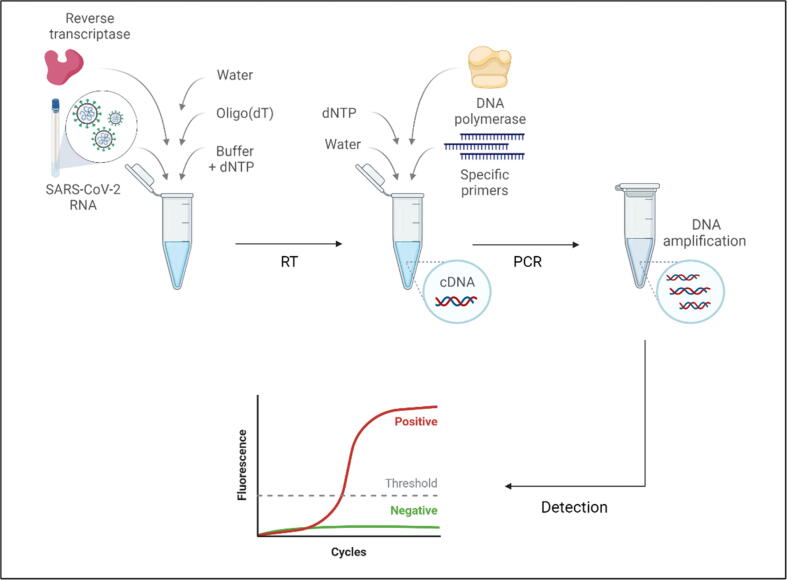
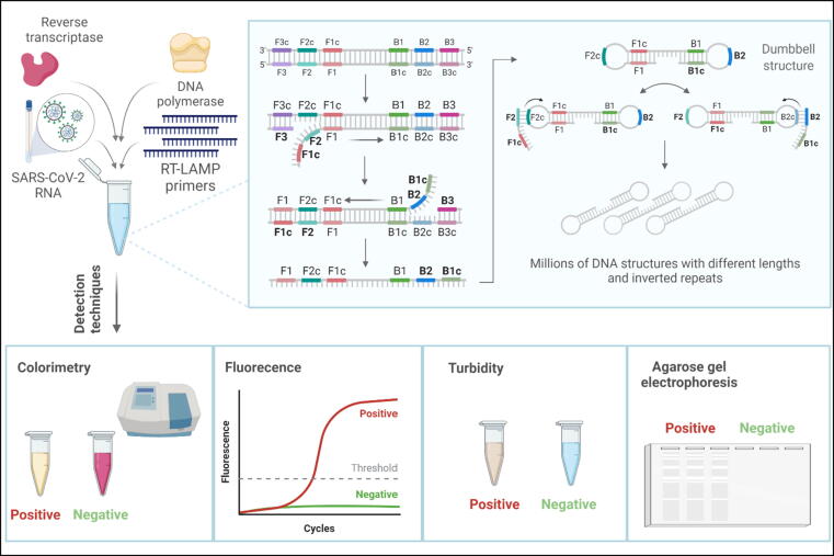
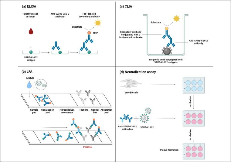
The ongoing novel COVID-19 has remained the center of attention, since its declaration as a pandemic in March 2020, due to its rapid and uncontrollable worldwide spread. Diagnostic tests are the first line of defense against the transmission of this infectious disease among individuals, with reverse-transcription quantitative polymerase chain reaction (RT-qPCR) being the approved gold standard for showing high sensitivity and specificity in detecting SARS-CoV-2. However, alternative tests are being invested due to the global demand for facilities, reagents, and healthcare workers needed for rapid population-based testing. Also, the rapid evolution of the viral genome and the emergence of new variants necessitates updating the existing methods. Scientists are aiming to improve tests to be affordable, simple, fast, and at the same time accurate, and efficient, as well as friendly user testing. The current diagnostic methods are either molecular-based that detect nucleic acids abundance, like RT-qPCR and reverse-transcription loop-mediated isothermal amplification (RT-LAMP); or immunologically based that detect the presence of antigens or antibodies in patients' specimens, like enzyme-linked immunosorbent assay (ELISA), lateral flow assay (LFA), chemiluminescent immunoassay (CLIA), and neutralization assay. In addition to these strategies, sensor-based or CRISPR applications are promising tools for the rapid detection of SARS-CoV-2. This review summarizes the most recent updates on the SARS-CoV-2 detection methods with their limitations. It will guide researchers, epidemiologists, and clinicians in identifying a more rapid, reliable, and sensitive method of diagnosing SARS-CoV-2 including the most recent variant of concern Omicron.

Keywords: COVID-19; Detection methods; RT-qPCR; SARS-CoV-2; Serology; Specimens; Variants.


Conflict of interest statement

The authors declare that they have no known competing financial interests or personal relationships that could have appeared to influence the work reported in this paper.

Figures

Fig. 1
Fig. 1
(a) SARS-CoV-2 structural proteins and their genomic locations. (b) The genome organization of the viral genome residing nonstructural proteins (nsp) and structural protein-encoding genes (S, E, M, and N). nsp12, translated by open reading frame (ORF1ab), encodes for RNA-dependent RNA polymerase.
Fig. 2
Fig. 2
Reverse transcription quantitative polymerase chain reaction (RT-qPCR) basic steps. Complementary DNA (cDNA) is first synthesized by preparing a master mix containing an RNA template and reverse transcriptase enzyme. Then, another master mix that includes gene-specific primers and the enzyme DNA polymerase is added to initiate the PCR reaction, resulting in millions of DNA targeted sequence copies. Real-time fluorescence detection shows the amplification curve for positive samples.
Fig. 3
Fig. 3
Reverse transcription loop-mediated isothermal amplification (RT-LAMP) single-tube reaction contains the RNA template, four primers, reverse transcriptase, and DNA polymerase enzymes. In this reaction, forward inner primer (FIP), forward outer primer (F3), backward inner primer (BIP), and backward outer primer (B3) bind to their complementary regions on the targeted DNA sequence (cDNA). New strands are synthesized afterward by DNA polymerase enzyme, in which complementary sequences cause the formation of dumbbell structures. Further amplifications result in millions of DNA inverted repeats with different lengths that can be detected by techniques like colorimetric, fluorescence, turbidity, and agarose gel electrophoresis.
Fig. 4
Fig. 4
Serological methods to detect SARS-CoV-2 or anti-SARS-CoV-2 antibodies from patients' samples. (a) Enzyme-linked immunosorbent assay (ELISA) utilizes SARS-CoV-2 antigen immobilized on wells, and antibodies from a blood or serum sample will form antigen–antibody complexes. After washing unbound antibodies, a secondary antibody labeled with horseradish peroxidase (HRP) is added with its substrate to produce a color resulting from binding to the primary antibody. (b) Lateral flow assay (LFA) detects SARS-CoV-2 antigens in infected individuals by running specimens through the sample pad to the conjugation pad, where antigens bind to specific and non-specific conjugated antibodies. The complexes flow through the nitrocellulose membrane to the test line to bind to anti-SARS-CoV-2 antibodies and produce color, indicating a positive sample. The control line producing a color designates successful analyte flow. (c) Chemiluminescence immunoassay (CLIA) has SARS-CoV-2 antigen-conjugated magnetic beads immobilized on its surface. anti-SARS-CoV-2 antibody from a blood or serum sample binds to the antigen, which in turn, a secondary antibody conjugated with a luminescent molecule binds to the primary antibody. Eventually, a substrate is added to yield light production. (d) In neutralization assays, anti-SARS-CoV-2 antibodies and SARS-CoV-2 are added to the Vero E6 cell culture. This assay tests the antibodies’ ability to block the binding of the virus to cell receptors, thereby preventing plaque formation.

References

    1. Abduljalil J.M. Laboratory diagnosis of SARS-CoV-2: available approaches and limitations. New Microbes New Infect. 2020;36 - PMC - PubMed
    1. Abdullah M., Sudrajat D.G., Muzellina V.N., Kurniawan J., Rizka A., Utari A.P., Pribadi R.R., Idrus M.F., Yusra Y., Meilany S., Surandy A., Shatri H., Rinaldi I., Pitoyo C.W., Renaldi K. The value of anal swab RT-PCR for COVID-19 diagnosis in adult Indonesian patients. BMJ Open Gastroenterol. 2021;8 - PMC - PubMed
    1. Abe, K. T., Li, Z., Samson, R., Samavarchi-Tehrani, P., Valcourt, E.J., Wood, H., Budylowski, P., Dupuis, A.P., 2ND, Girardin, R.C., Rathod, B., Wang, J.H., Barrios-Rodiles, M., Colwill, K., Mcgeer, A.J., Mubareka, S., Gommerman, J.L., Durocher, Y., Ostrowski, M., Mcdonough, K.A., Drebot, M.A., Drews, S.J., Rini, J.M., Gingras, A.C., 2020. A simple protein-based surrogate neutralization assay for SARS-CoV-2. JCI Insight, 5. - PMC - PubMed
    1. Abid S.A., Ahmed Muneer A., Al-Kadmy I.M.S., Sattar A.A., Beshbishy A.M., Batiha G.E., Hetta H.F. Biosensors as a future diagnostic approach for COVID-19. Life Sci. 2021;273 - PMC - PubMed
    1. Aithal S., Mishriki S., Gupta R., Sahu R.P., Botos G., Tanvir S., Hanson R.W., Puri I.K. SARS-CoV-2 detection with aptamer-functionalized gold nanoparticles. Talanta. 2022;236 - PMC - PubMed

LinkOut - more resources