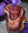
The Role of the Trapezius in Stabilization of the Acromioclavicular Joint: A Biomechanical Evaluation
- PMID: 36186709
- PMCID: PMC9520165
- DOI: 10.1177/23259671221118943
The Role of the Trapezius in Stabilization of the Acromioclavicular Joint: A Biomechanical Evaluation
Abstract
Background: Acromioclavicular joint (ACJ) injuries are common, and many are adequately treated nonoperatively. Biomechanical studies have mainly focused on static ligamentous stabilizers. Few studies have quantified ACJ stabilization provided by the trapezius.
Purpose/hypothesis: To elucidate the stabilization provided by the trapezius to the ACJ during scapular internal and external rotation (protraction and retraction). It was hypothesized that sequential trapezial resection would result in increasing ACJ instability.
Study design: Controlled laboratory study.
Methods: A biomechanical approach was pursued, with 10 cadaveric shoulders with the trapezius anatomically force loaded to normal. The trapezius was then serially transected over 8 trials, which alternated between clavicular defects (CD) and scapular defects (SD); each sequential defect consisted of 25% of the clavicular or scapular trapezial attachment. After each defect, specimens were tested with angle-controlled scapular internal and external rotation (12°) with rotary torque measurements to evaluate ACJ stability.
Results: The mean resistance in rotary torque for 12° of scapular internal rotation (protraction) with native specimens was 7.0 ± 2.0 N·m. Overall, internal rotation demonstrated a significant decrease in ACJ stability with trapezial injury (P < .001). Eight sequential defects resulted in the following significant percentage decreases in rotary torque from native internal rotation: 1.5% (25% CD; 0% SD), 5.6% (25% CD; 25% SD), 5.1% (50% CD; 25% SD), 6.5% (50% CD; 50% SD), 3.8% (75% CD; 50% SD), 7.1% (75% CD; 75% SD), 6.7% (100% CD; 75% SD), and 12.3% (100% CD 100% SD) (P < .001). The mean resistance in rotary torque for 12° of scapular external rotation (retraction) with native specimens was 7.1 ± 1.7 N·m. External rotation did not demonstrate a significant decrease in ACJ stability with trapezial injury (P = .596). The 8 sequential defects resulted in decreases in rotary torque from native external rotation of 0%, 3.8%, 4.0%, 3.2%, 3.5%, 3.4%, 4.2%, and 0.7%.
Conclusion: Trapezial injury resulted in increased instability in the setting of scapular internal rotation (protraction) of the ACJ.
Clinical relevance: These findings validate the inclusion of deltotrapezial fascial injury consideration in the modified Rockwood classification system. Repair of the trapezial insertion on the ACJ may provide improved outcomes in the setting of ACJ reconstruction.
Keywords: acromioclavicular joint; biomechanics; dynamic stabilizer; shoulder; trapezius.
© The Author(s) 2022.
Conflict of interest statement
One or more of the authors has declared the following potential conflict of interest or source of funding: A.D.M. has received consulting fees from Arthrex and Astellas Pharma, speaking fees from Arthrex and Kairos Surgical, and honoraria from Arthrosurface. AOSSM checks author disclosures against the Open Payments Database (OPD). AOSSM has not conducted an independent investigation on the OPD and disclaims any liability or responsibility relating thereto.
Figures


References
-
- Beitzel K, Sablan N, Chowaniec DM, et al. Sequential resection of the distal clavicle and its effects on horizontal acromioclavicular joint translation. Am J Sports Med. 2012;40(3):681–685. - PubMed
-
- Carofino BC, Mazzocca AD. The anatomic coracoclavicular ligament reconstruction: surgical technique and indications. J Shoulder Elbow Surg. 2010;19(2)(suppl):37–46. - PubMed
-
- Cerciello S, Berthold DP, Uyeki C, et al. Anatomic coracoclavicular ligament reconstruction (ACCR) using free tendon allograft is effective for chronic acromioclavicular joint injuries at mid-term follow-up. Knee Surg Sports Traumatol Arthrosc. 2021;29(7):2096–2102. - PubMed
-
- Debski RE, Parsons IM III, Fenwick J, Vangura A. Ligament mechanics during three degree-of-freedom motion at the acromioclavicular joint. Ann Biomed Eng. 2000;28(6):612–618. - PubMed
-
- Debski RE, Parsons IM IV, Woo SL, Fu FH. Effect of capsular injury on acromioclavicular joint mechanics. J Bone Joint Surg Am. 2001;83(9):1344–1351. - PubMed
LinkOut - more resources
Full Text Sources

