

Corneal imaging with blue-light optical coherence microscopy
- PMID: 36187260
- PMCID: PMC9484440
- DOI: 10.1364/BOE.465707
Corneal imaging with blue-light optical coherence microscopy
Abstract
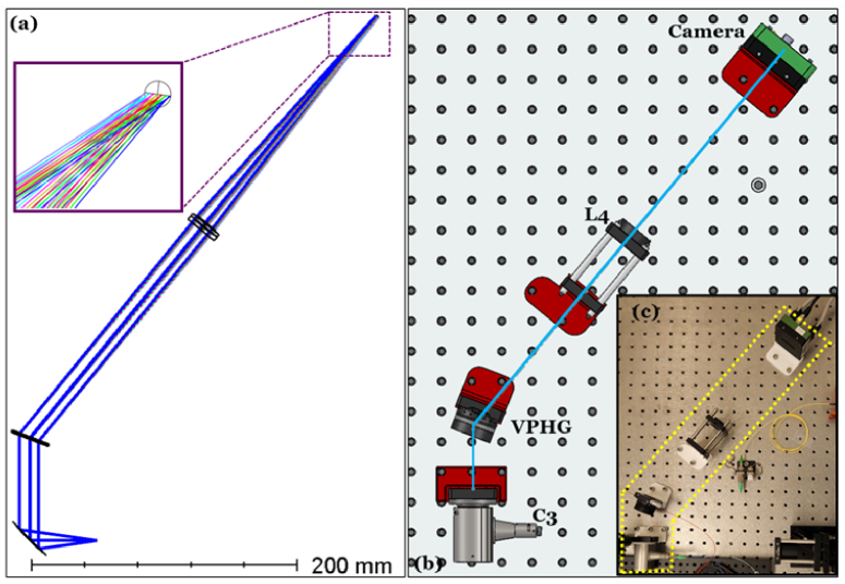
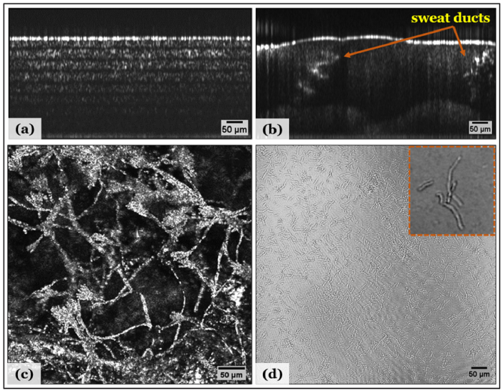
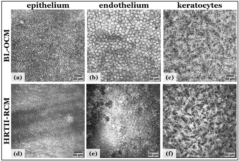
Corneal imaging is important for the diagnostic and therapeutic evaluation of many eye diseases. Optical coherence tomography (OCT) is extensively used in ocular imaging due to its non-invasive and high-resolution volumetric imaging characteristics. Optical coherence microscopy (OCM) is a technical variation of OCT that can image the cornea with cellular resolution. Here, we demonstrate a blue-light OCM as a low-cost and easily reproducible system to visualize corneal cellular structures such as epithelial cells, endothelial cells, keratocytes, and collagen bundles within stromal lamellae. Our blue-light OCM system achieved an axial resolution of 12 µm in tissue over a 1.2 mm imaging depth, and a lateral resolution of 1.6 µm over a field of view of 750 µm × 750 µm.
© 2022 Optica Publishing Group under the terms of the Optica Open Access Publishing Agreement.
Conflict of interest statement
David Huang: Optovue Inc. (F, I, P, R). These potential conflicts of interest have been reviewed and managed by OHSU. Other authors declare no relevant conflicts of interest related to this article.
Figures


References
-
- Flaxman S. R., Bourne R. R. A., Resnikoff S., Ackland P., Braithwaite T., Cicinelli M. V., Das A., Jonas J. B., Keeffe J., Kempen J. H., Leasher J., Limburg H., Naidoo K., Pesudovs K., Silvester A., Stevens G. A., Tahhan N., Wong T. Y., Taylor H. R., Vision S., Loss Expert Group of the Global Burden of Disease , “Global causes of blindness and distance vision impairment 1990-2020: a systematic review and meta-analysis,” Lancet Glob Health 5(12), e1221–e1234 (2017).10.1016/S2214-109X(17)30393-5 - DOI - PubMed
LinkOut - more resources
Full Text Sources