

Clinicopathological characterization of ten patients with primary malignant melanoma of the esophagus and literature review
- PMID: 36187400
- PMCID: PMC9516654
- DOI: 10.4251/wjgo.v14.i9.1739
Clinicopathological characterization of ten patients with primary malignant melanoma of the esophagus and literature review
Abstract
Background: Primary malignant melanoma of the esophagus (PMME) is a rare malignant disease and has not been well characterized in terms of clinicopathology and survival.
Aim: To investigate the clinical features and survival factors in Chinese patients with PMME.
Methods: The clinicopathological findings of ten cases with PMME treated at Henan Provincial People's Hospital were summarized. Moreover, the English- and Chinese-language literature that focused on Chinese patients with PMME from 1980 to September 2021 was reviewed and analyzed. Univariate and multivariate analyses were employed to investigate the clinicopathologic factors that might be associated with survival.
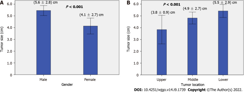
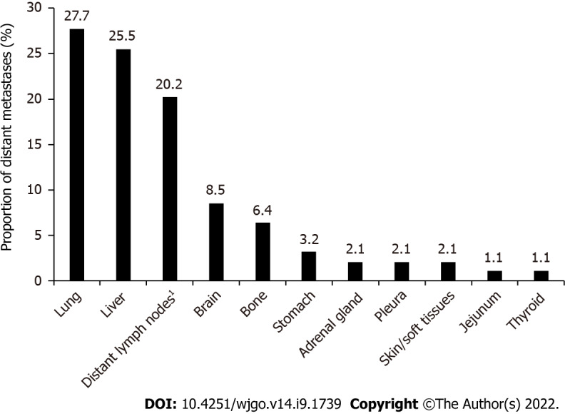
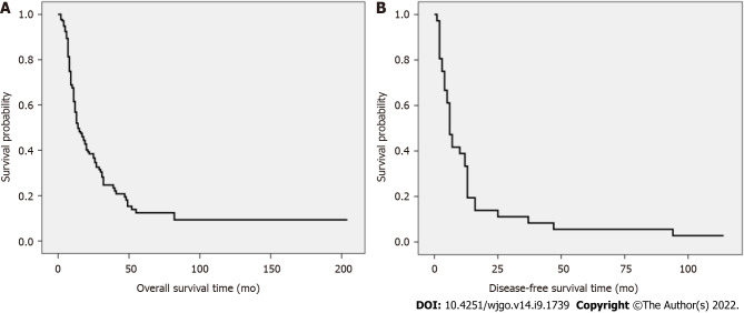
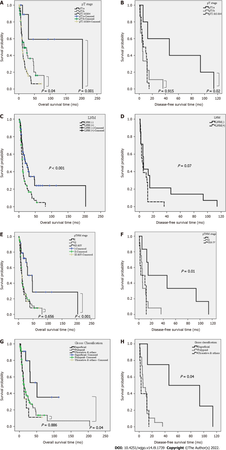
Results: A total of 290 Chinese patients with PMME, including ten from our hospital and 280 from the literature were enrolled in the present study. Only about half of the patients (55.8%) were accurately diagnosed before surgery. Additionally, 91.1% of the patients received esophagectomy, and 88 patients (36.5%) received adjuvant therapy after surgery. The frequency of lymph node metastasis (LNM) was 51.2% (107/209), and LNM had a positive rate of 45.3% even when the tumor was confined to the submucosal layer. The risk of LNM increased significantly with the pT stage [P < 0.001, odds ratio (OR): 2.47, 95% confidence interval (CI): 1.72-3.56] and larger tumor size (P = 0.006, OR: 1.21, 95%CI: 1.05-1.38). The median overall survival (OS) was 11.0 mo (range: 1-204 mo). The multivariate Cox analysis showed both the pT stage [P = 0.005, hazard ratio (HR): 1.70, 95%CI: 1.17-2.47] and LNM (P = 0.009, HR: 1.78, 95%CI: 1.15-2.74) were independent prognostic factors for OS. The median disease-free survival (DFS) was 5.3 mo (range: 0.8-114.1 mo). The multivariate analysis indicated that only the advanced pT stage (P = 0.02, HR: 1.93, 95%CI: 1.09-3.42) was a significant independent indicator of poor RFS in patients with PMME.
Conclusion: The correct diagnosis of PMME before surgery is low, and physicians should pay more attention to avoid a misdiagnosis or missed diagnosis. Extended lymph node dissection should be emphasized in surgery for PMME even though the tumor is confined to the submucosal layer. Both the LNM and pT stage are independent prognosis factors for OS, and the pT stage is the prognosis factor for DFS in patients with PMME.
Keywords: Clinicopathological characteristics; Primary malignant melanoma of the esophagus; Recurrence; Survival; Treatment.
©The Author(s) 2022. Published by Baishideng Publishing Group Inc. All rights reserved.
Conflict of interest statement
Conflict-of-interest statement: All the authors report no relevant conflicts of interest for this article.
Figures
References
-
- Lam AK. Updates on World Health Organization classification and staging of esophageal tumors: implications for future clinical practice. Hum Pathol. 2021;108:100–112. - PubMed
-
- Lam KY, Law S, Wong J. Malignant melanoma of the oesophagus: clinicopathological features, lack of p53 expression and steroid receptors and a review of the literature. Eur J Surg Oncol. 1999;25:168–172. - PubMed
-
- Schizas D, Mylonas KS, Bagias G, Mastoraki A, Ioannidi M, Kanavidis P, Hasemaki N, Karavokyros I, Theodorou D, Liakakos T. Esophageal melanoma: a systematic review and exploratory recurrence and survival analysis. Dis Esophagus. 2019 - PubMed
-
- Dai L, Wang ZM, Xue ZQ, He M, Yuan Y, Shang XQ, Chen KN Chinese Cooperative Primary Malignant Melanoma of the Esophagus Group (CCPMMEG) Results of surgical treatment for primary malignant melanoma of the esophagus: A multicenter retrospective study. J Thorac Cardiovasc Surg. 2020 - PubMed
LinkOut - more resources
Full Text Sources
