

Heterotaxy Syndrome with Polysplenia, Fused Adrenal Glands, and Diabetes Mellitus
- PMID: 36187466
- PMCID: PMC9520153
- DOI: 10.1177/11795468221116851
Heterotaxy Syndrome with Polysplenia, Fused Adrenal Glands, and Diabetes Mellitus
Abstract
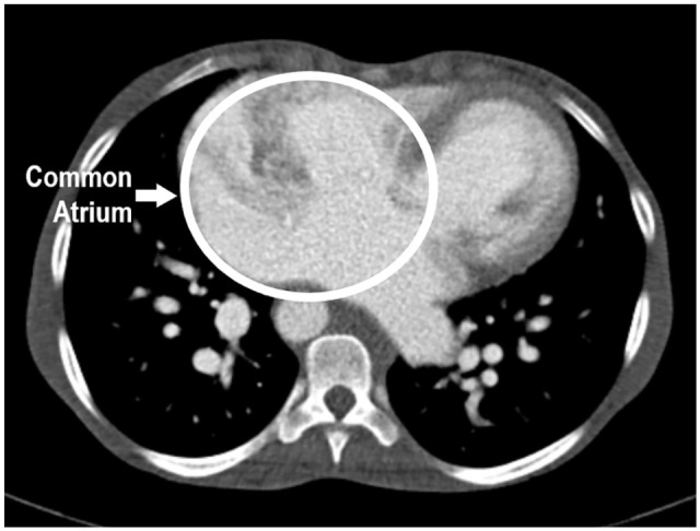
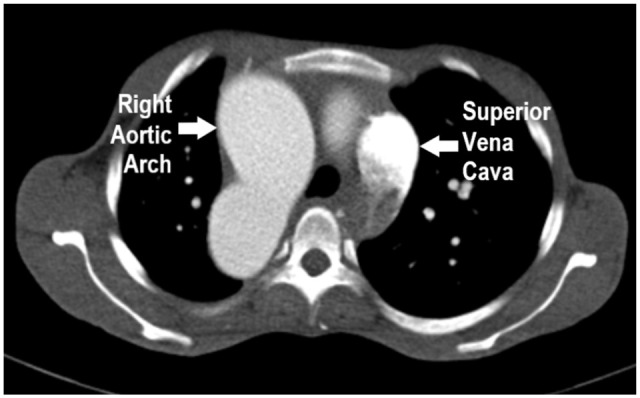
Heterotaxy syndrome is a rare congenital heart disease with a disarrangement of the heart and abdominal organs. We present a young African female with features of heart failure, diffuse irregular cardiac murmurs, and palpable, tender epigastric mass. A chest and abdominal computed tomography (CT) identified heterotaxy syndrome with left isomerism and fused adrenal glands. This case highlights the feature of fused adrenal glands in a patient with polysplenia.
Keywords: Congenital heart disease; diabetes mellitus; fused adrenal glands; imaging; polysplenia; pulmonary arterial hypertension.
© The Author(s) 2022.
Conflict of interest statement
Declaration of Conflicting Interests: The author(s) declared no potential conflicts of interest with respect to the research, authorship, and/or publication of this article.
Figures


Similar articles
-
Heterotaxy polysplenia syndrome in an adult female with complete endocardial cushion defect.Radiol Case Rep. 2021 Feb 24;16(5):1080-1084. doi: 10.1016/j.radcr.2021.02.015. eCollection 2021 May. Radiol Case Rep. 2021. PMID: 33717387 Free PMC article.
-
Polysplenia syndrome with duodenal and pancreatic dysplasia in a Holstein calf: a case report.BMC Vet Res. 2017 Sep 29;13(1):292. doi: 10.1186/s12917-017-1213-2. BMC Vet Res. 2017. PMID: 28962659 Free PMC article.
-
Incidental Finding of Heterotaxy Syndrome in a Patient With Pulmonary Embolism: A Case Report and Concise Review.Cureus. 2022 Apr 20;14(4):e24326. doi: 10.7759/cureus.24326. eCollection 2022 Apr. Cureus. 2022. PMID: 35607583 Free PMC article.
-
Dorsal pancreas agenesis and polysplenia/heterotaxy syndrome: a novel association with aortic coarctation and a review of the literature.JOP. 2007 Jul 9;8(4):433-7. JOP. 2007. PMID: 17625295 Review.
-
The nomenclature, definition and classification of cardiac structures in the setting of heterotaxy.Cardiol Young. 2007 Sep;17 Suppl 2:1-28. doi: 10.1017/S1047951107001138. Cardiol Young. 2007. PMID: 18039396 Review.
References
-
- Jacobs JP, Anderson RH, Weinberg PM, et al.. The nomenclature, definition and classification of cardiac structures in the setting of heterotaxy. Cardiol Young. 2007;17 Suppl 2:1-28. - PubMed
-
- Yim D, Nagata H, Lam CZ, et al.. Disharmonious patterns of heterotaxy and isomerism: how often are the classic patterns breached? Circ Cardiovasc Imaging. 2018;11:e006917-e006919. - PubMed
-
- Loomba RS, Nijhawan K, Anderson R. Impact of era, type of isomerism, and ventricular morphology on survival in heterotaxy. World J Pediatr Congenit Heart Surg. 2016;7:54-62. - PubMed
-
- Ticho BS, Goldstein AM, Van Praagh R. Extracardiac anomalies in the heterotaxy syndromes with focus on anomalies of midline-associated structures. Am J Cardiol. 2000;85:729-734. - PubMed
Publication types
LinkOut - more resources
Full Text Sources
