. 2022 Sep 14:13:1003999.
doi: 10.3389/fphys.2022.1003999. eCollection 2022.

Piezo buffers mechanical stress via modulation of intracellular Ca2+ handling in the Drosophila heart

Affiliations

Piezo buffers mechanical stress via modulation of intracellular Ca2+ handling in the Drosophila heart

Luigi Zechini et al. Front Physiol. .

Abstract

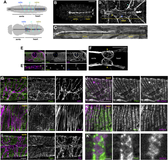
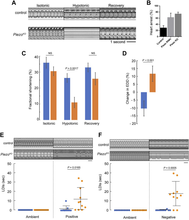
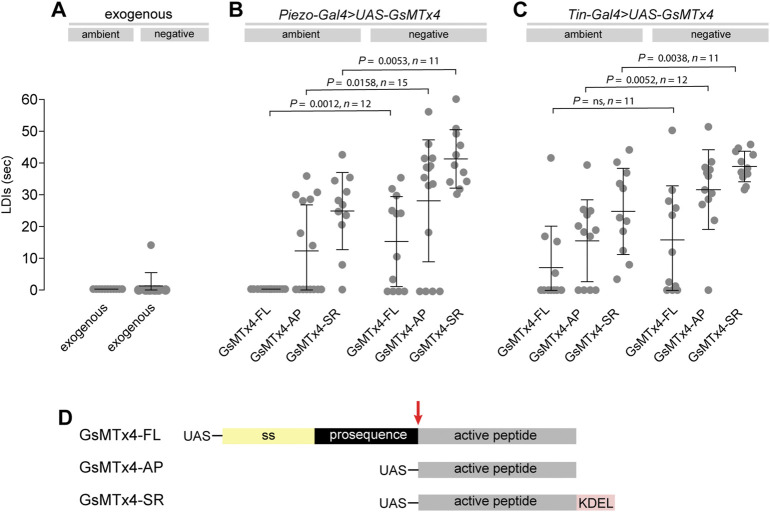
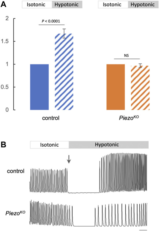
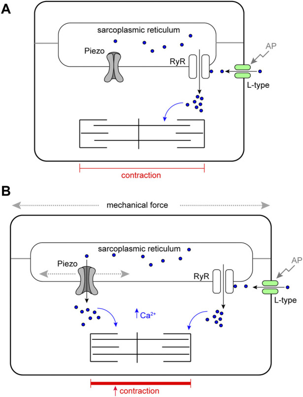
Throughout its lifetime the heart is buffeted continuously by dynamic mechanical forces resulting from contraction of the heart muscle itself and fluctuations in haemodynamic load and pressure. These forces are in flux on a beat-by-beat basis, resulting from changes in posture, physical activity or emotional state, and over longer timescales due to altered physiology (e.g. pregnancy) or as a consequence of ageing or disease (e.g. hypertension). It has been known for over a century of the heart's ability to sense differences in haemodynamic load and adjust contractile force accordingly (Frank, Z. biology, 1895, 32, 370-447; Anrep, J. Physiol., 1912, 45 (5), 307-317; Patterson and Starling, J. Physiol., 1914, 48 (5), 357-79; Starling, The law of the heart (Linacre Lecture, given at Cambridge, 1915), 1918). These adaptive behaviours are important for cardiovascular homeostasis, but the mechanism(s) underpinning them are incompletely understood. Here we present evidence that the mechanically-activated ion channel, Piezo, is an important component of the Drosophila heart's ability to adapt to mechanical force. We find Piezo is a sarcoplasmic reticulum (SR)-resident channel and is part of a mechanism that regulates Ca2+ handling in cardiomyocytes in response to mechanical stress. Our data support a simple model in which Drosophila Piezo transduces mechanical force such as stretch into a Ca2+ signal, originating from the SR, that modulates cardiomyocyte contraction. We show that Piezo mutant hearts fail to buffer mechanical stress, have altered Ca2+ handling, become prone to arrhythmias and undergo pathological remodelling.

Keywords: Drosophila; Frank-Starling; Piezo; calcium; heart; mechanosensitive ion channel; mechanotransduction; sacroplasmic recticulum.


Conflict of interest statement

The authors declare that the research was conducted in the absence of any commercial or financial relationships that could be construed as a potential conflict of interest.

Figures

FIGURE 1
FIGURE 1
Piezo expression in the Drosophila heart. (A) Schematic figure of a Drosophila larva (upper) and adult (lower) depicting the heart, aorta and associated cell types. Cardiomyocytes (dark grey), ostia (blue), intracardiac valves (yellow), and alary muscles (al, cyan) are shown. Conical chamber is indicated with dashed line. (B) Piezo expression in a late stage Drosophila embryo revealed by in situ hybridization chain reaction. (C) Piezo expression in a larva revealed by Piezo-Gal4>UAS-Piezo::GFP. (D) Piezo expression in an adult revealed by Piezo-Gal4>UAS-Piezo::GFP. Conical chamber is indicated with dashed line. (E) Piezo-Gal4>UAS-nRFP (green), seven up-LacZ (blue, bracket in E′) and actin (magenta) reveal Piezo expression in cardiomyocytes but not ostia. (F) Piezo expression in larval intracardiac valves (yellow arrowheads) revealed by Piezo-Gal4>UAS-Piezo::GFP. (G,H) Piezo subcellular localisation revealed by Piezo-Gal4>UAS-Piezo::GFP (green) in the aorta (G) and heart (H) in relation to actin filaments (magenta). (I) Piezo subcellular localisation revealed by Piezo-Gal4>UAS-Piezo::GFP (green) in the aorta relative to T-tubule marker anti-Discs large (Dlg, magenta). (J) Piezo subcellular localisation revealed by Piezo-Gal4>UAS-Piezo::GFP (green) in the heart relative to the SR marker (ER-tracker, magenta). (K) Piezo subcellular localisation revealed by Piezo-Gal4>UAS-Piezo::GFP (green) in the heart relative to the SR marker anti-Serca (magenta). (K′) shows a higher magnification view of the box marked in (K). Scale bars = 100 µm (B, E′), 200 µm (C,D), 50 µm (E,F), 20 μm, (G,H), 10 µm (I–K), 2 µm (K′). For all images anterior is to the left.
FIGURE 2
FIGURE 2
Piezo hearts fail to buffer mechanical stress. (A) M-mode kymograph traces for control (upper) and Piezo KO (lower) adult hearts in isotonic, hypotonic and isotonic (recovery) solutions. (B) Percentage of cardiac arrest during hypotonic stretch in control, Piezo-RNAi and Piezo KO animals. (n = 28, 23, and 24). (C) Fractional shortening in the conical chamber under isotonic (left), hypotonic (middle) and isotonic (recovery) conditions for control (blue, n = 12) and Piezo KO (orange, n = 12) hearts. There is no statistical difference in FS for control hearts in isotonic versus hypotonic solution (p = 0.19, n = 12, one-way ANOVA) (D) Response of the hearts to hypotonic stress. Percentage change in end diastolic diameter (EDD) for control (blue) and Piezo KO (orange) hearts measured at the conical chamber (n = 10 and 10). (E) Graph shows quantification of long diastolic intervals (LDIs) for control (blue, n = 11) and Piezo KO (orange, n = 11) hearts under conditions of ambient and positive (+220 mmHg) pressure. Each data point represents an individual heart preparation; the y-axis shows additive time in LDI over a 60 s period for each individual preparation. Typical M-mode kymograph trace for control and Piezo KO adult hearts are shown (upper panel). (F) Graph shows quantification of LDIs for control (blue, n = 10) and Piezo KO (orange, n = 10) hearts under conditions of ambient and negative (−482 mmHg) pressure. Each data point represents an individual heart preparation; the y-axis shows additive time in LDI over a 60 s period for each individual preparation. Typical M-mode kymograph trace for control and Piezo KO adult hearts are shown (upper panel). p-values, unpaired t-test. NS = not significant. Scale bar in A, E, F = 1 s.
FIGURE 3
FIGURE 3
Pharmacological inhibition of Piezo causes heart arrest under mechanical stress. (A–C) Quantification of LDIs with GsMTx-4 applied exogenously (A, 5mM; n = 12), or when expressed from a transgenic construct using Piezo-Gal4 (B) or Tin-Gal4 (C), under conditions of ambient and negative (−482 mmHg) pressure. Each data point represents an individual heart preparation; the y-axis shows additive time in LDI over a 60 s period for each individual preparation. p-values, unpaired t-test. (D) Schematic drawing of the GsMTx-4 transgenic constructs used in the study: secreted (GsMTx4-FL); non-secreted (GsMTx4-AP); SR-targeted (GsMTx4-SR). UAS = Upstream Activation Sequence; ss = signal sequence; KDEL = ER/SR translocation signal; red arrow = mature peptide cleavage site.
FIGURE 4
FIGURE 4
Elevated Ca2+ transients in response to hypotonic stretch require Piezo. (A) Summary data comparing Ca2+ transient amplitude in response to hypotonic stretch in control (blue) or Piezo KO (orange) cardiomyocytes (block colour = isotonic, stippling = hypotonic). Approximately 20 Ca2+ transients were measured in isotonic media to calculate mean amplitude per cardiomyocyte. The amplitudes of first 10 transients in hypotonic media were averaged. The resulting value was normalised to the isotonic value to allow comparison between genotypes. n = 18 (control) and 19 (Piezo KO ) individual cardiomyocytes from independent heart preparations. (B) Representative Ca2+ traces from control (upper) and Piezo KO (lower) cardiomyocytes in isotonic/hypotonic solution (arrow marks the change from isotonic to hypotonic solution). Time is on x-axis, fluorescence (arbitrary units) on y-axis, scale bar = 1 s. p values, unpaired t-test. NS = not significant.
FIGURE 5
FIGURE 5
Dilation of the heart lumen in Piezo KO . (A) Adult heart stained with phalloidin to reveal morphology. Heart length (yellow dashed line) and lumen cross-sectional area at the position of the 2nd abdominal segment (yellow solid line) were measured. Anterior is to left. (B) Heart length of control (blue) and Piezo KO (orange) adult hearts (n = 12; n = 12). (C) Optical x-z projection of control or Piezo KO hearts stained with phalloidin from first instar larva (left), 3-day old adult (middle) and 10-day old hypertensive adult (right). (D) Lumenal area in control (blue) and Piezo KO (orange) hearts for first instar larvae (n = 7; n = 7), normotensive adults (n = 12; n = 11) and hypertensive adults (n = 12; n = 12). (E) Cardiomyocyte nuclear area in control (blue) and Piezo KO (orange) adult hearts under normotensive (n = 42; n = 45) and hypertensive conditions (n = 43; n = 41) n = number of cardiomyocytes from at least 3 individual hearts. (F) Control (left) and 10-day old hypertensive adult (right); in the hypertensive fly the heart is exposed to high blood pressure in the grossly swollen haemolymph-filled abdomen (white arrow indicates swollen abdomen). p-values, unpaired t-test.
FIGURE 6
FIGURE 6
Schematic model of Piezo in the Drosophila cardiomyocyte. (A) Cardiomyocyte contraction is controlled by cytoplasmic Ca2+. Depolarisation of the membrane by an action potential (AP) opens sarcolemmal L-type voltage-gated Ca2+ channels activating further Ca2+ release from the sarcoplasmic reticulum (SR) through Ryanodine Receptors (RyR). Cytoplasmic Ca2+ activates the myofilaments and leads to contraction of the cell. In circumstances where mechanical force is within normal range, SR localised Piezo channels are closed. (B) In circumstances where mechanical force (e.g. stretch or compression). exceeds the normal range SR, localised Piezo channels open, additional Ca2+ is released from the SR elevating the cytoplasmic Ca2+ pool, this leads to a stronger contraction.

References

    1. Adler C. P., Friedburg H., Herget G. W., Neuburger M., ScHwalb H. (1996). Variability of cardiomyocyte DNA content, ploidy level and nuclear number in mammalian hearts. Virchows Arch. 429 (2-3), 159–164. 10.1007/BF00192438 - DOI - PubMed
    1. Adler C. P., Friedburg H. (1986). Myocardial DNA content, ploidy level and cell number in geriatric hearts: Post-mortem examinations of human myocardium in old age. J. Mol. Cell. Cardiol. 18 (1), 39–53. 10.1016/s0022-2828(86)80981-6 - DOI - PubMed
    1. Ahmad S. M. (2017). Conserved signaling mechanisms in Drosophila heart development. Dev. Dyn. 246 (9), 641–656. 10.1002/dvdy.24530 - DOI - PMC - PubMed
    1. Alayari N. N., Vogler G., Taghli-Lamallem O., Ocorr K., Bodmer R., Cammarato A. (2009). Fluorescent labeling of Drosophila heart structures. J. Vis. Exp. 32, 1423. 10.3791/1423 - DOI - PMC - PubMed
    1. Alcaino C., Knutson K., Gottlieb P. A., Farrugia G., Beyder A. (2017). Mechanosensitive ion channel Piezo2 is inhibited by D-GsMTx4. Channels (Austin) 11 (3), 245–253. 10.1080/19336950.2017.1279370 - DOI - PMC - PubMed

LinkOut - more resources