

Magnetic resonance imaging features of canine intradural/extramedullary intervertebral disc extrusion in seven cases
- PMID: 36187811
- PMCID: PMC9517942
- DOI: 10.3389/fvets.2022.1003042
Magnetic resonance imaging features of canine intradural/extramedullary intervertebral disc extrusion in seven cases
Abstract
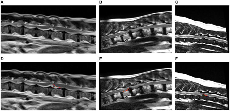
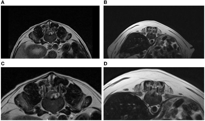
Intervertebral disc disease, including intervertebral disc extrusions and protrusions, is the most common spinal cord disorder in dogs. Atypical and uncommon intervertebral disc herniations include intradural/intramedullary disc extrusion, intervertebral foraminal disc extrusion and intervertebral disc herniation (Schmorl's node). Intradural/extramedullary disc extrusion is the least common type of intervertebral disc herniation in veterinary medicine, characterized by extruded disc material within the intradural space. To date, only one study has been published in veterinary medicine reporting intradural/extramedullary disc extrusions. In this study, low field MRI was used, and the authors could not find any MRI features to diagnose with confidence an intradural/extramedullary disc location of the extruded disc material. The aim of this study was to describe the high field (1.5T) MRI characteristics of surgically confirmed intradural/extramedullary disc extrusions. This is a retrospective, multicentric and descriptive study. Inclusion criteria was surgical confirmation of intradural/extramedullary disc extrusion by durotomy and complete MRI study of the spine. Seven cases were included. Images were reviewed by a radiology resident and a certified radiologist, with emphasis on the following signs: "Golf-tee sign" (widening of the subarachnoid space cranial and caudal to the lesion), "Beak sign" (pointed and sharp compressive lesion) and "Y sign" (division of the dura and arachnoid layers). MRI showed a "Y sign" in all the cases (7/7) seen from the T2-weighted sagittal views, while "Golf-tee sign" was not recognized in any of the cases (0/7). Additionally, "beak sign" was present in half of the cases (4/7). "Y sign" maybe a reliable MRI feature for identifying intradural/medullary disc extrusions from the MRI study. As the arachnoid is peeled from the dura by the disc herniation there is a splitting of the arachnoid mater and the ventral dura. The intradural disc material will be surrounded by CSF signal intensity margin, giving the appearance of a Y, which can be identified from the T2-weighted sagittal images.
Keywords: MRI; Y sign; disc extrusion; dog; extramedullary; intradural.
Copyright © 2022 Casado, Fernandes, Lourinho, Gonçalves, Clark, Violini and Carrera.
Conflict of interest statement
The authors declare that this study received funding from Linnaeus Veterinary Limited. The funder was not involved in the study design, collection, analysis, interpretation of data, the writing of this article, or the decision to submit it for publication.
Figures
Similar articles
-
Thoracolumbar intradural disc herniation in eight dogs: clinical, low-field magnetic resonance imaging, and computed tomographic myelography findings.Vet Radiol Ultrasound. 2015 Mar-Apr;56(2):160-7. doi: 10.1111/vru.12213. Epub 2014 Sep 26. Vet Radiol Ultrasound. 2015. PMID: 25263808
-
The specific sagittal magnetic resonance imaging of intradural extra-arachnoid lumbar disc herniation.Case Rep Med. 2012;2012:383451. doi: 10.1155/2012/383451. Epub 2012 Feb 16. Case Rep Med. 2012. PMID: 22431932 Free PMC article.
-
Intradural Disc a Diagnostic Dilemma: Case Series and Review of Literature.Asian J Neurosurg. 2018 Oct-Dec;13(4):1033-1036. doi: 10.4103/ajns.AJNS_55_17. Asian J Neurosurg. 2018. PMID: 30459862 Free PMC article.
-
Intradural disc herniation at the L2/3 level: a case report and literature review.Ann Palliat Med. 2022 Sep;11(9):3005-3013. doi: 10.21037/apm-22-1017. Ann Palliat Med. 2022. PMID: 36217628 Review.
-
A Review of Fibrocartilaginous Embolic Myelopathy and Different Types of Peracute Non-Compressive Intervertebral Disk Extrusions in Dogs and Cats.Front Vet Sci. 2015 Aug 18;2:24. doi: 10.3389/fvets.2015.00024. eCollection 2015. Front Vet Sci. 2015. PMID: 26664953 Free PMC article. Review.
Cited by
-
Case Report: Dural infiltration of intervertebral disc material mimicking spinal cord meningioma in a dog.Front Vet Sci. 2025 Jul 1;12:1621529. doi: 10.3389/fvets.2025.1621529. eCollection 2025. Front Vet Sci. 2025. PMID: 40666731 Free PMC article.
-
Case report: Magnetic resonance imaging features with postoperative improvement of atypical cervical glioma characterized by predominant extramedullary distribution in a dog.Front Vet Sci. 2024 May 22;11:1400139. doi: 10.3389/fvets.2024.1400139. eCollection 2024. Front Vet Sci. 2024. PMID: 38840642 Free PMC article.
-
Case series: Cervical far-lateral and combined cervical far lateral/foraminal intervertebral disk extrusions in 10 dogs.Front Vet Sci. 2024 Nov 6;11:1465182. doi: 10.3389/fvets.2024.1465182. eCollection 2024. Front Vet Sci. 2024. PMID: 39606656 Free PMC article.
References
-
- Priester WA. Canine intervertebral disc disease — occurrence by age, breed, and sex among 8,117 cases. Theriogenology. (1976) 6:293–303. 10.1016/0093-691X(76)90021-2 - DOI
-
- Bergknut N, Auriemma E, Wijsman S, Voorhout G, Hagman R, Lagerstedt A-S, et al. . Evaluation of intervertebral disk degeneration in chondrodystrophic and nonchondrodystrophic dogs by use of Pfirrmann grading of images obtained with low-field magnetic resonance imaging. Am J Vet Res. (2011) 72:893–8. 10.2460/ajvr.72.7.893 - DOI - PubMed
LinkOut - more resources
Full Text Sources
Research Materials