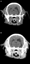
Case report: Atypical and chronic masticatory muscle myositis in a 5-month old Cavalier King Charles Spaniel. Clinical and diagnostic findings, treatment and successful outcome
- PMID: 36187837
- PMCID: PMC9516294
- DOI: 10.3389/fvets.2022.955758
Case report: Atypical and chronic masticatory muscle myositis in a 5-month old Cavalier King Charles Spaniel. Clinical and diagnostic findings, treatment and successful outcome
Abstract
Masticatory muscle myositis (MMM) is the second most common inflammatory myopathy diagnosed in dogs, but it is rarely described in puppies. The disease is characterized by the production of autoantibodies against 2M myofibers contained in masticatory muscle, although the cause of this production is still unclear. The aim of the present case report was to describe the clinical presentation, diagnostic findings, treatment, and follow-up of an atypical case of chronic masticatory muscle myositis in a very young dog. A 5-month old Cavalier king Charles Spaniel (CKCS) was presented to the AniCura Istituto Veterinario Novara with a two weeks, progressive history of lethargy and difficulty in food prehension. Neurological examination revealed bilateral masticatory muscle atrophy, mandibular ptosis with the jaw kept open, inability to close the mouth without manual assistance, jaw pain, and bilateral reduction of palpebral reflex and menace reaction; vision was maintained. A myopathy was suspected. Computed tomography (CT), magnetic resonance imaging (MRI), enzyme-linked immunosorbent assay test for 2M antibodies, and histopathological examination of masticatory muscle biopsy confirmed the diagnosis of MMM. Glucocorticoids treatment was started and clinical signs promptly improved. To the authors' knowledge, this is the first case describing mandibular ptosis in a dog affected by chronic MMM, successfully managed with medical treatment and the first report describing the CT and MRI findings in a young CKCS affected by MMM.
Keywords: CKCS; CT; MRI; dog; masticatory muscle myositis.
Copyright © 2022 Di Tosto, Callegari, Matiasek, Lacava, Salvatore, Muñoz Declara, Betti and Tirrito.
Conflict of interest statement
The authors declare that the research was conducted in the absence of any commercial or financial relationships that could be construed as a potential conflict of interest.
Figures


References
-
- Castejon-Gonzalez AC, Soltero-Rivera M, Brown DC, Reiter AM. Treatment outcome of 22 dogs with masticatory muscle myositis (1999–2015). J Vet Dent. (2018) 35:281–9. 10.1177/0898756418813536 - DOI
-
- Melmed C, Shelton GD, Bergman R, Barton C. Masticatory muscle myositis: pathogenesis, diagnosis, and treatment. Compendium Continuing Edu Practis Vet North Am Ed. (2004) 26:590–605. - PubMed
Publication types
LinkOut - more resources
Full Text Sources

