

Cross-species recognition and molecular basis of SARS-CoV-2 and SARS-CoV binding to ACE2s of marine animals
- PMID: 36187898
- PMCID: PMC9517163
- DOI: 10.1093/nsr/nwac122
Cross-species recognition and molecular basis of SARS-CoV-2 and SARS-CoV binding to ACE2s of marine animals
Abstract
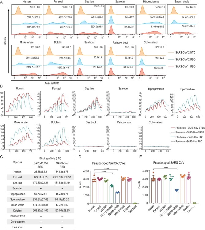
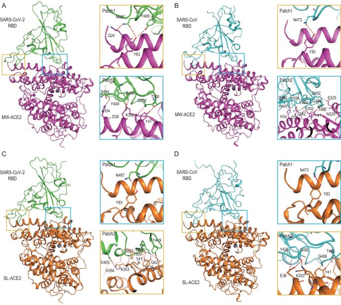
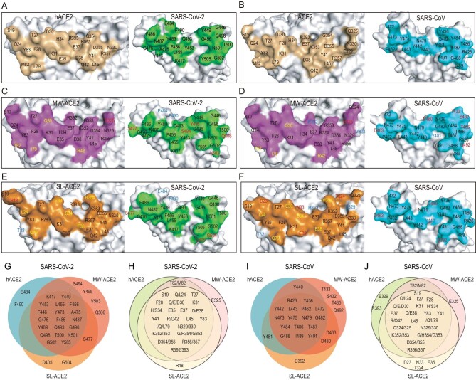
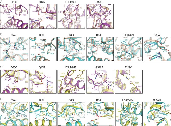
Severe acute respiratory syndrome coronavirus 2 (SARS-CoV-2) has an extremely broad host range that includes hippopotami, which are phylogenetically closely related to whales. The cellular ACE2 receptor is one of the key determinants of the host range. Here, we found that ACE2s from several marine mammals and hippopotami could efficiently bind to the receptor-binding domain (RBD) of both SARS-CoV and SARS-CoV-2 and facilitate the transduction of SARS-CoV and SARS-CoV-2 pseudoviruses into ACE2-expressing cells. We further resolved the cryo-electron microscopy complex structures of the minke whale ACE2 and sea lion ACE2, respectively, bound to the RBDs, revealing that they have similar binding modes to human ACE2 when it comes to the SARS-CoV-2 RBD and SARS-CoV RBD. Our results indicate that marine mammals could potentially be new victims or virus carriers of SARS-CoV-2, which deserves further careful investigation and study. It will provide an early warning for the prospective monitoring of marine mammals.
Keywords: SARS-CoV-2; cross-species recognition; cryo-EM structure; marine animals.
© The Author(s) 2022. Published by Oxford University Press on behalf of China Science Publishing & Media Ltd.
Figures
Similar articles
-
Molecular basis of hippopotamus ACE2 binding to SARS-CoV-2.J Virol. 2024 May 14;98(5):e0045124. doi: 10.1128/jvi.00451-24. Epub 2024 Apr 9. J Virol. 2024. PMID: 38591877 Free PMC article.
-
Broad host range of SARS-CoV-2 and the molecular basis for SARS-CoV-2 binding to cat ACE2.Cell Discov. 2020 Sep 29;6:68. doi: 10.1038/s41421-020-00210-9. eCollection 2020. Cell Discov. 2020. PMID: 33020722 Free PMC article.
-
Molecular Basis of Mink ACE2 Binding to SARS-CoV-2 and Its Mink-Derived Variants.J Virol. 2022 Sep 14;96(17):e0081422. doi: 10.1128/jvi.00814-22. Epub 2022 Aug 24. J Virol. 2022. PMID: 36000849 Free PMC article.
-
Mutations derived from horseshoe bat ACE2 orthologs enhance ACE2-Fc neutralization of SARS-CoV-2.PLoS Pathog. 2021 Apr 9;17(4):e1009501. doi: 10.1371/journal.ppat.1009501. eCollection 2021 Apr. PLoS Pathog. 2021. PMID: 33836016 Free PMC article.
-
Receptor recognition and cross-species infections of SARS coronavirus.Antiviral Res. 2013 Oct;100(1):246-54. doi: 10.1016/j.antiviral.2013.08.014. Epub 2013 Aug 29. Antiviral Res. 2013. PMID: 23994189 Free PMC article. Review.
Cited by
-
Sarbecovirus RBD indels and specific residues dictating multi-species ACE2 adaptiveness.Nat Commun. 2024 Oct 14;15(1):8869. doi: 10.1038/s41467-024-53029-3. Nat Commun. 2024. PMID: 39402048 Free PMC article.
-
Structural basis for receptor binding and broader interspecies receptor recognition of currently circulating Omicron sub-variants.Nat Commun. 2023 Jul 21;14(1):4405. doi: 10.1038/s41467-023-39942-z. Nat Commun. 2023. PMID: 37479708 Free PMC article.
-
Expression, purification and immunogenicity analyses of receptor binding domain protein of severe acute respiratory syndrome coronavirus 2 from delta variant.Vet Res Forum. 2024;15(12):657-663. doi: 10.30466/vrf.2024.2013858.4037. Epub 2024 Dec 15. Vet Res Forum. 2024. PMID: 39816631 Free PMC article.
-
Structural basis of increased binding affinities of spikes from SARS-CoV-2 Omicron variants to rabbit and hare ACE2s reveals the expanding host tendency.mBio. 2024 Feb 14;15(2):e0298823. doi: 10.1128/mbio.02988-23. Epub 2023 Dec 19. mBio. 2024. PMID: 38112468 Free PMC article.
-
Cryo-EM structures and binding of mouse and human ACE2 to SARS-CoV-2 variants of concern indicate that mutations enabling immune escape could expand host range.PLoS Pathog. 2023 Apr 5;19(4):e1011206. doi: 10.1371/journal.ppat.1011206. eCollection 2023 Apr. PLoS Pathog. 2023. PMID: 37018380 Free PMC article.
References
LinkOut - more resources
Full Text Sources
Miscellaneous