

A case series of eosinophilic myocarditis: different faces of the same coin
- PMID: 36187932
- PMCID: PMC9518670
- DOI: 10.1093/ehjcr/ytac388
A case series of eosinophilic myocarditis: different faces of the same coin
Abstract
Background: Eosinophilic myocarditis (EM) is a rare form of myocarditis with various aetiologies and dire consequences if not diagnosed and treated expeditiously.
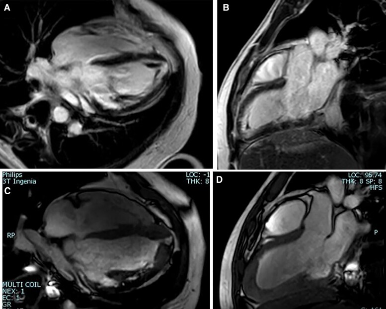
Case summary: We report three cases of EM at different stages of the disease with differing clinical manifestations. We highlight the diagnostic workup including the role of multimodality imaging and endomyocardial biopsy (EMB), and the treatment strategies.
Discussion: EM is an underdiagnosed and potentially life-threatening disease. Therefore, a high clinical suspicion for EM should arise when patients with signs and symptoms of cardiovascular disease develop hypereosinophilia or vice versa. Early identification of this condition using multimodality imaging and EMB is of paramount importance as the disease may progress to the irreversible late fibrotic stage if treatment is delayed.
Keywords: Case series; Eosinophilic myocarditis; Heart failure; Hypereosinophilia.
© The Author(s) 2022. Published by Oxford University Press on behalf of the European Society of Cardiology.
Figures


References
-
- Brambatti M, Matassini MV, Adler ED, Klingel K, Camici PG, Ammirati E. Eosinophilic myocarditis. J Am Coll Cardiol 2017;70:2363–2375. - PubMed
-
- Fozing T, Zouri N, Tost A, Breit R, Seeck G, Koch C, et al. . Management of a patient with eosinophilic myocarditis and normal peripheral eosinophil count case report and literature review. Circ Heart Fail 2014;7(4):692–694. - PubMed
-
- Parrillo JE, Borer JS, Henry WL, Wolff SM, Fauci AS. The cardiovascular manifestation of the hypereosinophilc syndrome. Prospective study of 26 patients, with review of the literature. Am J Med 1979;67:572–582. - PubMed
-
- Perazzolo Marra M, Thiene G, Rizzo S, De Lazzari M, Carturan E, Tona F, et al. . Cardiac magnetic resonance features of biopsy-proven endomyocardial diseases. JACC Cardiovasc Imaging 2014;7(3):309–312. - PubMed
Publication types
LinkOut - more resources
Full Text Sources