Case Reports
. 2022 Sep 19;17(11):4408-4412.
doi: 10.1016/j.radcr.2022.08.067. eCollection 2022 Nov.

Idiopathic pneumatosis cystoides coli: An uncommon cause of pneumoperitoneum

Affiliations
Case Reports

Idiopathic pneumatosis cystoides coli: An uncommon cause of pneumoperitoneum

André Peixoto et al. Radiol Case Rep. .

Abstract

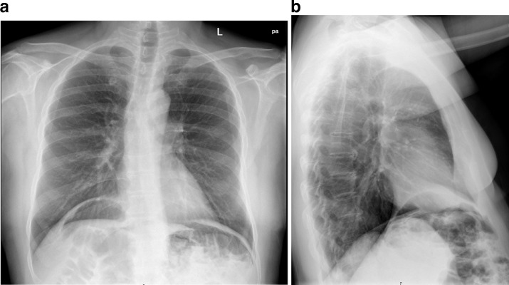
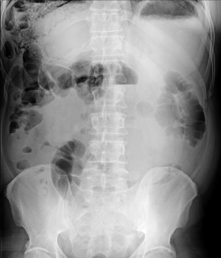
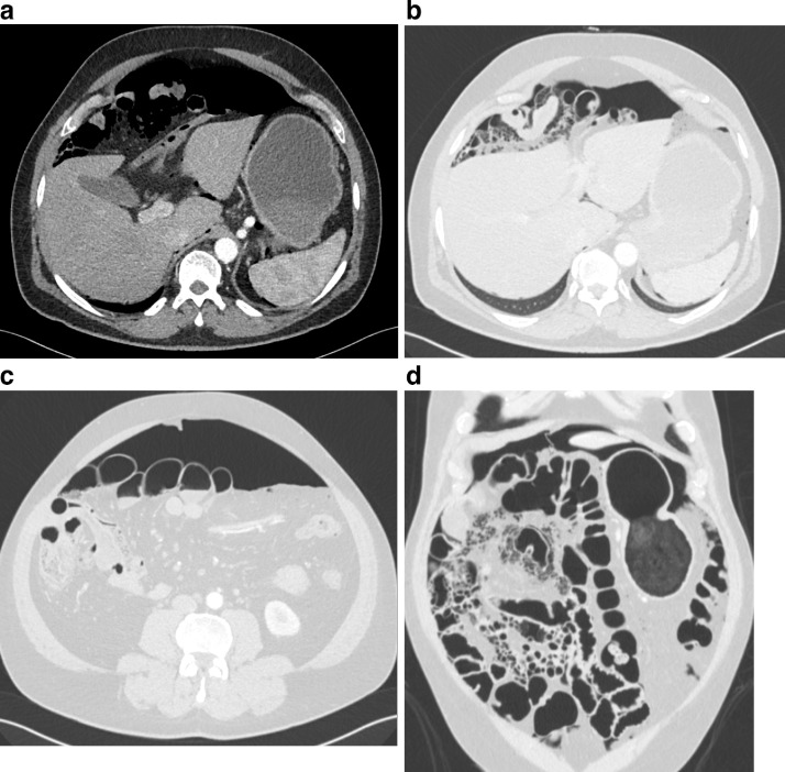
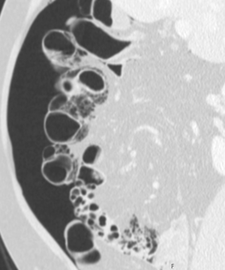
Pneumatosis cystoides intestinalis (PCI) is a rare entity characterized by the presence of gaseous cystic within the intestinal wall. The primary or idiopathic type represents 15% of cases and is a self-limited or chronic benign entity. The secondary type represents 85% of cases and is associated with various factors, such as surgery, pharmacotherapy, chemotherapy, autoimmune diseases, inflammatory diseases, and pulmonary illness. Pneumatosis cystoides intestinalis affects the colon (pneumatosis cystoides coli) in about half of the cases. The differential diagnosis of PCI includes potentially life-threatening diseases that cause pneumatosis intestinalis. The misdiagnosis of PCI is common and can lead to unnecessary treatments and surgical procedures. We describe an asymptomatic pneumoperitoneum incidentally seen on chest radiograph. The cause was pneumatosis cystoides coli, which did not require treatment.

Keywords: Pneumatosis cystoides coli; Pneumatosis cystoides intestinalis; Pneumatosis intestinalis; Pneumoperitoneum.


Figures

Fig 1
Fig. 1
Chest radiograph, posteroanterior (A) and lateral (B) views show bilateral subdiaphragmatic free air (pneumoperitoneum). In the upper abdominal quadrant, along the intestine's wall, gas pockets radiolucency is noted. There were no signs of pulmonary infection.
Fig 2
Fig. 2
Old abdominal radiograph demonstrates multiple gas pockets radiolucency, in grape-like cluster or honeycomb-shaped appearance, along the intestine's wall, predominantly in the right upper abdominal quadrant.
Fig 3
Fig. 3
Enhanced computerized tomography, axial (A-C) and coronal (D) images show pneumoperitoneum and multiple intestinal air cysts within the colon's wall, with a diffuse distribution, more numerous on the right colon.
Fig 4
Fig. 4
Follow-up enhanced computerized tomography, sagittal image continued to demonstrate pneumoperitoneum and colon with gas-filled cysts on the subserosa and submucosa.

Similar articles

Cited by

References

    1. Wang YJ, Wang YM, Zheng YM, Jiang HQ, Zhang J. Pneumatosis cystoides intestinalis: six case reports and a review of the literature. BMC Gastroenterol. 2018;18(1):100. doi: 10.1186/s12876-018-0794-y. - DOI - PMC - PubMed
    1. Ksiadzyna D, Peña AS. Segmental pneumatosis cystoides coli: computed tomography-facilitated diagnosis. Rev Esp Enferm Dig. 2016;108(8):510–513. doi: 10.17235/reed.2015.3790/2015. - DOI - PubMed
    1. Wu LL, Yang YS, Dou Y, Liu QS. A systematic analysis of pneumatosis cystoids intestinalis. World J Gastroenterol. 2013;19(30):4973–4978. doi: 10.3748/wjg.v19.i30.4973. - DOI - PMC - PubMed
    1. Ribolla M, Conti L, Baldini E, Palmieri G, Grassi C, Banchini F, et al. Asymptomatic pneumoperitoneum in pneumatosis coli: a misleading operative indication. Int J Surg Case Rep. 2020;69:92–95. doi: 10.1016/j.ijscr.2020.03.042. - DOI - PMC - PubMed
    1. Greenstein AJ, Nguyen SQ, Berlin A, Corona J, Lee J, Wong E, et al. Pneumatosis intestinalis in adults: management, surgical indications, and risk factors for mortality. J Gastrointest Surg. 2007;11(10):1268–1274. doi: 10.1007/s11605-007-0241-9. - DOI - PubMed

Publication types

LinkOut - more resources