

Annular pancreas: Radiological features of a rare case of infantile vomiting
- PMID: 36188077
- PMCID: PMC9520511
- DOI: 10.1016/j.radcr.2022.08.063
Annular pancreas: Radiological features of a rare case of infantile vomiting
Abstract
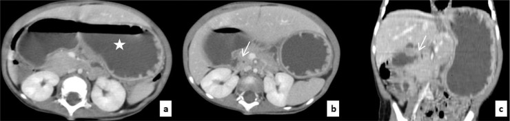
Our purpose is to illustrate the radiological aspects of the annular pancreas as an etiology of duodenal obstruction in infants. We report the case of a 4-month-old girl, who was admitted to our department with postprandial vomiting evolving since birth. The initial examination found a severely dehydrated patient. Abdominal ultrasound showed gross dilatation of the stomach and duodenum, it also showed pancreatic tissue surrounding the duodenum, suggesting a diagnosis of annular pancreas as the cause of the duodenal obstruction. Post-contrast abdominal CT showed the gastric and duodenal dilatation, and a ring of pancreatic tissue surrounding uncompletly the second portion of the duodenum. The patient underwent a bypass surgery which consisted in a duodeno-duodenostomy with simple post-operative follow-up and no recurrence of digestive symptoms. Annular pancreas is a rare pathology to be sought in neonatal obstruction. A good knowledge of radiological semiology is essential for a good diagnostic approach. However, surgery is the only effective way to diagnose and treat this pathology.
Keywords: Annular pancreas; Imaging; Pediatrics; Vomiting.
© 2022 Published by Elsevier Inc. on behalf of University of Washington.
Figures


Similar articles
-
Delayed presentation of duodenal diaphragm and annular pancreas in a 10-year-old girl: Case report.Radiol Case Rep. 2023 Nov 3;19(1):264-267. doi: 10.1016/j.radcr.2023.10.003. eCollection 2024 Jan. Radiol Case Rep. 2023. PMID: 38028306 Free PMC article.
-
A case of annular pancreas in a male adult.Fukushima J Med Sci. 2004 Dec;50(2):75-81. doi: 10.5387/fms.50.75. Fukushima J Med Sci. 2004. PMID: 15779573
-
A rare cause of proximal intestinal obstruction in adults - annular pancreas: a case report.Pan Afr Med J. 2011;10:56. Epub 2011 Dec 18. Pan Afr Med J. 2011. PMID: 22384302 Free PMC article.
-
Annular pancreas in neonates; Case series and review of literatures.Afr J Paediatr Surg. 2022 Apr-Jun;19(2):97-101. doi: 10.4103/ajps.AJPS_180_20. Afr J Paediatr Surg. 2022. PMID: 35017379 Free PMC article. Review.
-
Annular pancreas combined with distal stenosis. A report of four cases and review of the literature.Pediatr Med Chir. 2004 Jul-Aug;26(4):256-9. Pediatr Med Chir. 2004. PMID: 16366413 Review.
Cited by
-
Factors Associated with Postoperative Complications After Congenital Duodenal Obstruction Surgery: A Retrospective Study.Medicina (Kaunas). 2024 Oct 21;60(10):1722. doi: 10.3390/medicina60101722. Medicina (Kaunas). 2024. PMID: 39459509 Free PMC article.
-
Annular Pancreas: Insights Into the Diagnosis of a Rare Anatomical Malformation in Pediatric Patients.Cureus. 2025 Jun 10;17(6):e85702. doi: 10.7759/cureus.85702. eCollection 2025 Jun. Cureus. 2025. PMID: 40551727 Free PMC article.
-
Antenatal Diagnosis of Complete Annular Pancreas With Postnatal Surgical Follow-Up: A Case Report.Cureus. 2024 Aug 26;16(8):e67837. doi: 10.7759/cureus.67837. eCollection 2024 Aug. Cureus. 2024. PMID: 39323684 Free PMC article.
References
Publication types
LinkOut - more resources
Full Text Sources
