

Enhanced Antioxidant Effects of Naringenin Nanoparticles Synthesized using the High-Energy Ball Milling Method
- PMID: 36188293
- PMCID: PMC9521026
- DOI: 10.1021/acsomega.2c04148
Enhanced Antioxidant Effects of Naringenin Nanoparticles Synthesized using the High-Energy Ball Milling Method
Abstract
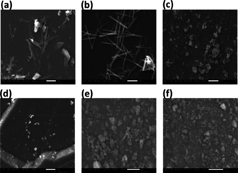
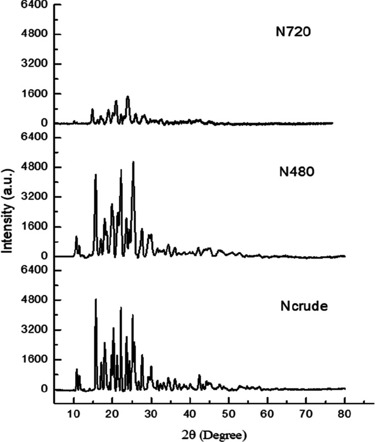
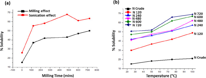
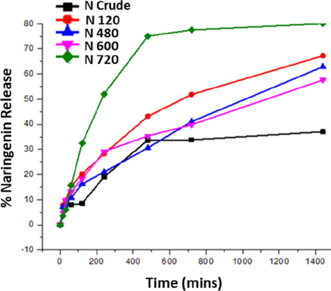
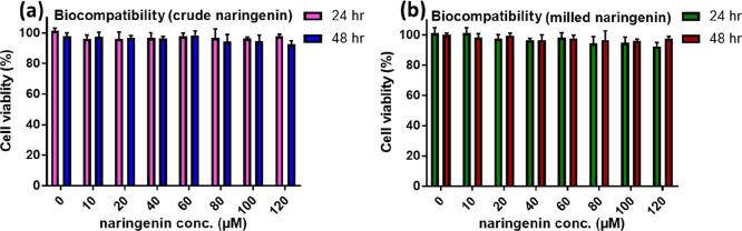
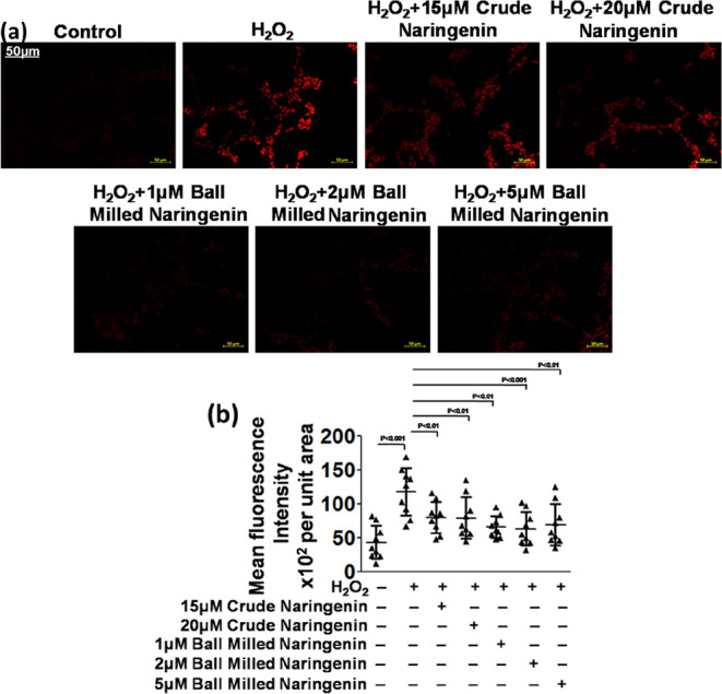
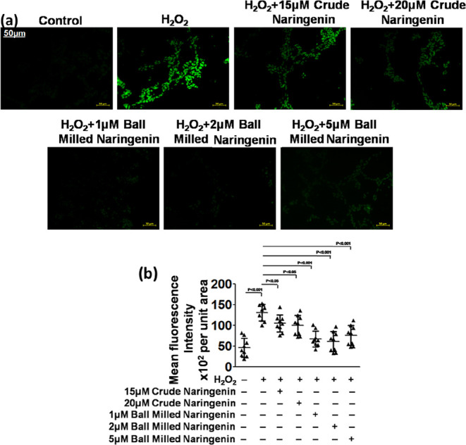
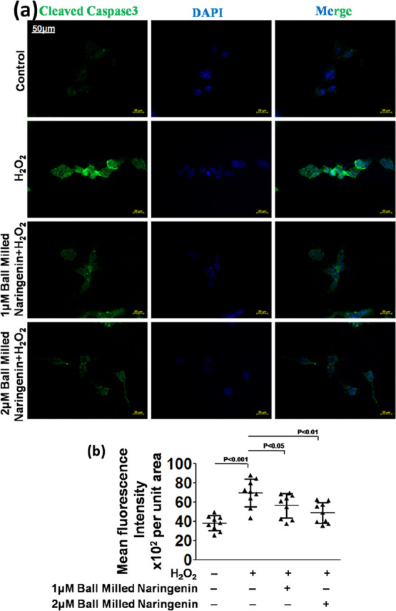
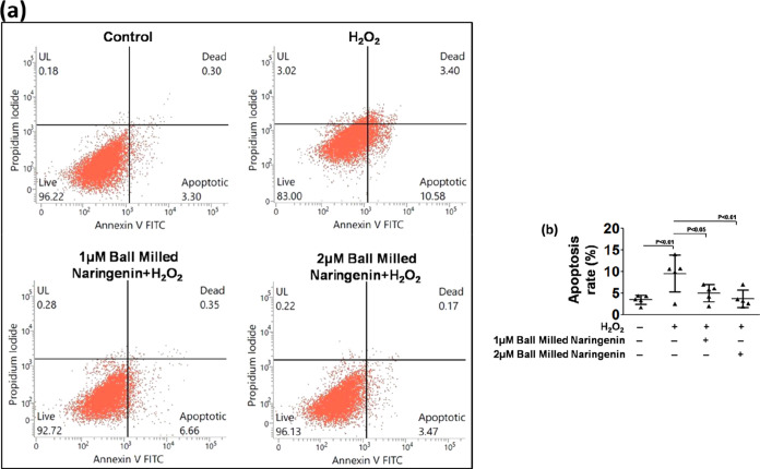
Naringenin, one of the flavonoid components, is majorly found in and obtained from grapefruits and oranges. Naringenin also acts as a potent antioxidant, which possesses hypolipidemic as well as anti-inflammatory potential. Naringenin reduces the expressions of several inflammatory mediators, viz., NF-κB, cycloxygenase-2, and other cytokine mediators. In spite of having various biological effects, the clinical application of naringenin is restricted due to its very poor aqueous solubility. In the present study, the high-energy ball milling method was employed for the preparation of naringenin nanoparticles without using any chemical with an aim to enhance the anti-oxidant potential of naringenin. The milled naringenin nanoparticles were characterized for their physicochemical properties using scanning electron microscopy (SEM) and X-ray diffraction. Additionally, the effects of milling time and temperature were further assessed on the solubility of crude and milled naringenin samples. The antioxidant potential of milled naringenin was evaluated with various assays such as DHE, DCFDA, and cleaved caspase-3 using SH-SY5Y human neuroblastoma cells. The nanoparticle size of naringenin after milling was confirmed using SEM analysis. Crystalline peaks for milled and crude samples of naringenin also established that both the naringenin forms were in the crystalline form. The solubility of naringenin was enhanced depending on the milling time and temperature. Moreover, crude and milled naringenin were found to be cytocompatible up to doses of 120 μM each for the duration of 24 and 48 h. It was also observed that milled naringenin at the doses of 1, 2, and 5 μM significantly reduced the levels of reactive oxygen species (ROS) generated by H2O2 and exhibited superior ROS scavenging effects as compared to those of crude or un-milled forms of naringenin. Furthermore, milled naringenin at the doses of 1 and 2 μM inhibited H2O2-induced cell death, as shown by immunofluorescence staining of cleaved caspase-3 and Annexin-V PI flow cytometry analysis. Conclusively, it could be suggested that the size reduction of naringenin using high-energy ball milling techniques substantially enhanced the antioxidant potential as compared to naïve or crude naringenin, which may be attributed to its enhanced solubility due to reduced size.
© 2022 The Authors. Published by American Chemical Society.
Conflict of interest statement
The authors declare no competing financial interest.
Figures
References
-
- Nogrady B. How Nanotechnology Can Flick the Immunity Switch. Nature 2021, 595, S18–S19. 10.1038/d41586-021-01790-6. - DOI
-
- Phillips I. D. J. Induction of a Light Requirement for the Germination of Lettuce Seed by Naringenin, and Its Removal by Gibberellic Acid. Nature 1961, 192, 240–241. 10.1038/192240a0. - DOI
-
- Ahmad A.; Fauzia E.; Kumar M.; Mishra R. K.; Kumar A.; Khan M. A.; Raza S. S.; Khan R. Gelatin-Coated Polycaprolactone Nanoparticle-Mediated Naringenin Delivery Rescue Human Mesenchymal Stem Cells from Oxygen Glucose Deprivation-Induced Inflammatory Stress. ACS Biomater. Sci. Eng. 2019, 5, 683–695. 10.1021/acsbiomaterials.8b01081. - DOI - PubMed
LinkOut - more resources
Full Text Sources
Research Materials
Miscellaneous
