Review
. 2022 Sep 16:13:913704.
doi: 10.3389/fneur.2022.913704. eCollection 2022.

Endovascular treatment of aneurysms of the paraophthalmic segment of the internal carotid artery: Current status

Affiliations
Review

Endovascular treatment of aneurysms of the paraophthalmic segment of the internal carotid artery: Current status

Yiheng Wang et al. Front Neurol. .

Abstract

The paraophthalmic segment of the internal carotid artery (ICA) originates from the distal border of the cavernous ICA and terminates at the posterior communicating artery. Aneurysms arising from the paraophthalmic segment represent ~5-10% of intradural aneurysms. Due to the advent of endovascular treatment (EVT) techniques, specifically flow-diverting stents (FDSs), EVT has become a good option for these aneurysms. A literature review on EVT for paraophthalmic segment aneurysms is necessary. In this review, we discuss the anatomy of the paraophthalmic segment, classification of the paraophthalmic segment aneurysms, EVT principle and techniques, and prognosis and complications. EVT techniques for paraophthalmic segment aneurysms include coil embolization, FDSs, covered stents, and Woven EndoBridge devices. Currently, coiling embolization remains the best choice for ruptured paraophthalmic segment aneurysms, especially to avoid long-term antiplatelet therapy for young patients. Due to the excessive use of antiplatelet therapy, unruptured paraophthalmic segment aneurysms that are easy to coil should not be treated with FDS. FDS is appropriate for uncoilable or failed aneurysms. Other devices cannot act as the primary choice but can be useful auxiliary tools. Both coiling embolization and FDS deployment can result in a good prognosis for paraophthalmic segment aneurysms. The overall complication rate is low. Therefore, EVT offers promising treatments for paraophthalmic segment aneurysms. In addition, surgical clipping continues to be a good choice for paraophthalmic segment aneurysms in the endovascular era.

Keywords: aneurysm; endovascular treatment; internal carotid artery; paraophthalmic segment; review.


Conflict of interest statement

The authors declare that the research was conducted in the absence of any commercial or financial relationships that could be construed as a potential conflict of interest.

Figures

Figure 1
Figure 1
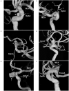
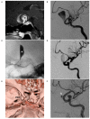
NYU paraophthalmic segment. (A,B) CTA (A) and DSA of the ICA (B) show the clinoidal and ophthalmic segments of the ICA. The segmentation is according to the classifications by Bouthillier and Ziyal. The asterisk in image (A) indicates the anterior clinoid process. (C) DSA shows the NYU segmentation. The clinoidal and ophthalmic segments of the ICA are combined into the paraophthalmic segment. CTA, computed tomography angiography; DSA, digital subtraction angiography; ICA, internal carotid artery; NYU, New York University; PcomA, posterior communicating artery.
Figure 2
Figure 2
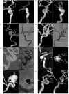
Classification of aneurysms of the paraophthalmic segment according to their locations. (A) Three-dimensional DSA shows a carotid cave aneurysm (arrow). (B) Three-dimensional DSA shows a clinoidal aneurysm (arrow) opposite the carotid cave. (C) Three-dimensional DSA shows an OphA aneurysm (arrow); the OphA was located away from the aneurysm neck. (D) Three-dimensional DSA shows an OphA aneurysm (arrow); the OphA arose from both the aneurysmal and ICA walls. (E) Three-dimensional DSA shows a suprasellar SHA aneurysm (arrow). (F) Three-dimensional DSA shows a paraclinoid SHA aneurysm (arrow). DSA, digital subtraction angiography; MCA, middle cerebral artery; OphA, ophthalmic artery; SHA, superior hypophyseal artery; PcomA, posterior communicating artery.
Figure 3
Figure 3
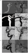
BBA and its differential diagnosis. (A) Left: Three-dimensional DSA of the ICA shows a mushroom-shaped BBA (arrow) on the paraophthalmic segment; Right: intraoperative image shows the fragile BBA without a definite neck (asterisk). (B) Left: DSA of the ICA shows an aneurysm (arrow) exactly like the BBA on the paraophthalmic segment; Right: intraoperative image shows that the aneurysm is saccular with a definite and stable neck (asterisk). BBA, blood blister-like aneurysm; DSA, digital subtraction angiography; ICA, internal carotid artery.
Figure 4
Figure 4
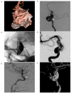
Other classifications of aneurysms of the paraophthalmic segment. (A) Three-dimensional DSA shows a fusiform aneurysm (arrow) of the paraophthalmic segment, which also belongs to dissection. (B) Three-dimensional DSA shows a large SHA aneurysm (arrow). (C) Three-dimensional DSA shows bilateral aneurysms: one is a coiled SHA aneurysm (arrow) and the other is a carotid cave aneurysm (arrowhead). (D) Three-dimensional DSA shows two tandem aneurysms: the large aneurysm is an SHA aneurysm (arrow), and the small aneurysm is a carotid cave aneurysm (arrowhead). DSA, Digital subtraction angiography; MCA, middle cerebral artery; OphA, ophthalmic artery; SHA, superior hypophyseal artery.
Figure 5
Figure 5
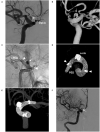
Various EVT techniques for aneurysms of the paraophthalmic segment. (A) Left, three-dimensional DSA shows a carotid artery aneurysm (arrow); right: Vaso CT shows the FDS covering the aneurysm. (B) Left, three-dimensional DSA shows two aneurysms (arrows) of the paraophthalmic segment; right: Vaso CT shows the FDS covering two aneurysms. (C) Left, three-dimensional DSA shows a clinoidal aneurysm (arrow); right: follow-up DSA shows that the aneurysms were coiled completely. (D) Left, three-dimensional DSA shows an OphA aneurysm (arrow); right: follow-up DSA shows that the aneurysm was coiled under stenting assistance. (E) Left, road map DSA shows a microcatheter into a paraclinoid-type SHA aneurysm (arrow); right: DSA shows the aneurysm was coiled. (F) Left, postoperative three-dimensional DSA shows a coiled suprasellar-type SHA aneurysm (arrow); right: Vaso CT shows the coils and stent. (G) Left, three-dimensional DSA shows a large OphA aneurysm (arrow); right: unsubtracted follow-up DSA shows that the aneurysm was coiled under stenting assistance. (H) Left, three-dimensional DSA shows OphA and SHA aneurysms (arrows); right: follow-up three-dimensional DSA shows that two aneurysms were coiled under stenting assistance. CT, computed tomography; DSA, digital subtraction angiography; EVT, endovascular treatment; FDS, flow-diverting stent; OphA, ophthalmic artery; SHA, superior hypophyseal artery; Vaso, vascular space occupancy.
Figure 6
Figure 6
EVT difficulty in aneurysms of the paraophthalmic segment. (A) Left, Road map navigation shows the microcatheter tip (arrow) positioned in the aneurysm sac with the assistance of stent semi-deployment. The stent sealed a part of the aneurysm neck and provided the opportunity for the microcrater to enter the aneurysm; right: unsubtracted DSA shows the coils in the aneurysm (arrow). (B) Left, three-dimensional DSA shows a wide-necked lobulated carotid cave aneurysm (arrow); right: DSA shows the loose packing of the coiling in the aneurysm because the microcatheter was knocked off the aneurysm, but contrast agent retention can be seen in the aneurysm (arrow). (C) Left, DSA shows a recanalized aneurysm due to low-density packing (arrow); right: follow-up DSA shows the aneurysm had a complete embolization (arrow) after repeated coiling. (D) Left, DSA shows a recanalized lobulated upper aneurysm (arrow) and a completely embolized lower aneurysm (arrowhead); right: X-ray film shows that the upper aneurysm had low-density packing due to difficult catheterization (arrow), and the lower aneurysm had satisfactory embolization (arrowhead). DSA, Digital subtraction angiography; EVT, endovascular treatment.
Figure 7
Figure 7
FDS deployment in a fusiform aneurysm under coiling assistance. (A,B) CTA (A) and DSA (B) show a large fusiform aneurysm (arrows) of the paraophthalmic segment. (C) X-ray shows the FDS deployment (PED) under the assistance of coiling (coils). (D) Postoperative DSA shows that the distal part of the aneurysm was coiled, and the proximal part (frame) was left. (E,F) Follow-up DSA at 6 months (E) and three-dimensional reconstructive DSA (F) show nearly complete occlusion; the arrows indicate the remnant neck. CTA, computed tomography angiography; DSA, digital subtraction angiography; FDS, flow-diverting stent; PED, pipeline embolization device.
Figure 8
Figure 8
FDS deployment in a giant aneurysm without coiling. (A) Sagittal MRI shows a giant aneurysm with jet flow in the middle (asterisk). (B) DSA shows the aneurysm sac. (C) X-ray shows that contrast agent retention (asterisk) can be seen after FDS (PED) deployment. (D) Postoperative DSA shows reduced blood flow into the aneurysm. (E) Postoperative 1-week CTA shows the location of the PED. (F) Follow-up DSA at 6 months shows that most of the aneurysm was occluded and that complete occlusion occurred, and the arrow indicates the remnant part. CTA, computed tomography angiography; DSA, digital subtraction angiography; FDS, flow-diverting stent; MRI, magnetic resonance imaging; PED, pipeline embolization device.
Figure 9
Figure 9
FDS deployment in a BBA with failed previous stent-assisted coiling. (A,B) Two-dimensional DSA (A) and three-dimensional DSA (B) show a BBA (arrows) with incomplete embolization of stent-assisted coiling. (C) Unsubtracted DSA shows the FDS (PED) crossing the BBA (arrow) and the FDS running in the stent (arrowheads). (D,E) Vaso CT without the vessel (D) and shows the FDS opened and covering the BBA in the stent (arrowheads). (F) Postoperative DSA shows the BBA without immediate complete occlusion; complete occlusion was expected by long-term follow-up. BBA, blister-like aneurysm; DSA, digital subtraction angiography; FDS, flow-diverting stent; PED, pipeline embolization device; Vaso, vascular space occupancy.

Similar articles

Cited by

References

    1. Gibo H, Lenkey C, Rhoton AL Jr. Microsurgical anatomy of the supraclinoid portion of the internal carotid artery. J Neurosurg. (1981) 55:560–74. 10.3171/jns.1981.55.4.0560 - DOI - PubMed
    1. Shapiro M, Becske T, Riina HA, Raz E, Zumofen D, Jafar JJ, et al. . Toward an endovascular internal carotid artery classification system. AJNR Am J Neuroradiol. (2014) 35:230–6. 10.3174/ajnr.A3666 - DOI - PMC - PubMed
    1. Bhogal P, Aguilar Perez M, Sauder G, Bazner H, Ganslandt O, Henkes H. Management of paraophthalmic aneurysms: review of endovascular treatment strategies. Ophthalmologe. (2018) 115:114–22. 10.1007/s00347-017-0497-8 - DOI - PubMed
    1. Wisniewski K, Tomasik B, Bobeff EJ, Stefanczyk L, Hupalo M, Jaskolski DJ. Predictors for ophthalmic segment aneurysms recanalization after coiling and flow diverter embolization in 6- and 12-month follow-up. J Clin Neurosci. (2019) 68:151–7. 10.1016/j.jocn.2019.07.007 - DOI - PubMed
    1. De Jesus O, Sekhar LN, Riedel CJ. Clinoid and paraclinoid aneurysms: surgical anatomy, operative techniques, and outcome. Surg Neurol. (1999) 51:477–87. 10.1016/S0090-3019(98)00137-2 - DOI - PubMed