. 2022 Jun;3(2):116-129.
doi: 10.1002/aac2.12049. Epub 2022 Apr 19.

Another Wrinkle with Age: Aged Collagen and Intra-peritoneal Metastasis of Ovarian Cancer

Affiliations

Another Wrinkle with Age: Aged Collagen and Intra-peritoneal Metastasis of Ovarian Cancer

Elizabeth I Harper et al. Aging Cancer. 2022 Jun.

Abstract

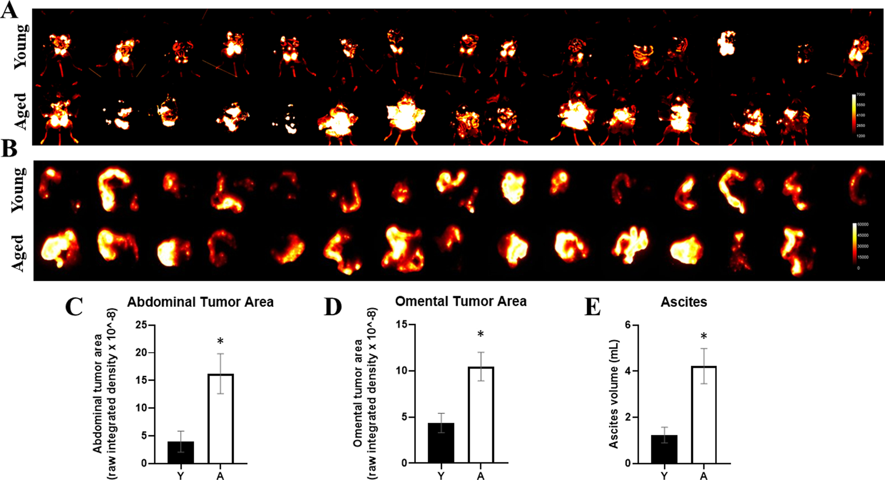
Background: Age is the most significant risk factor for ovarian cancer (OvCa), the deadliest gynecologic malignancy. Metastasizing OvCa cells adhere to the omentum, a peritoneal structure rich in collagen, adipocytes, and immune cells. Ultrastructural changes in the omentum and the omental collagen matrix with aging have not been evaluated.

Aim: The aim of this study was to test the hypothesis that age-related changes in collagen in the ovarian tumor microenvironment promote OvCa metastatic success in the aged host.

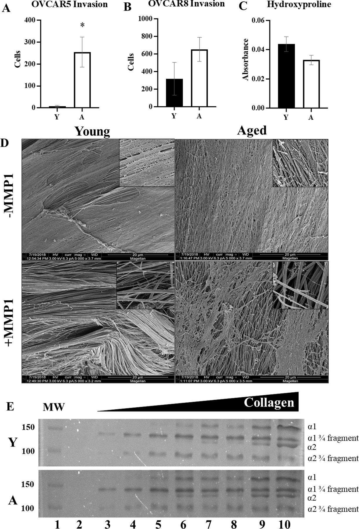
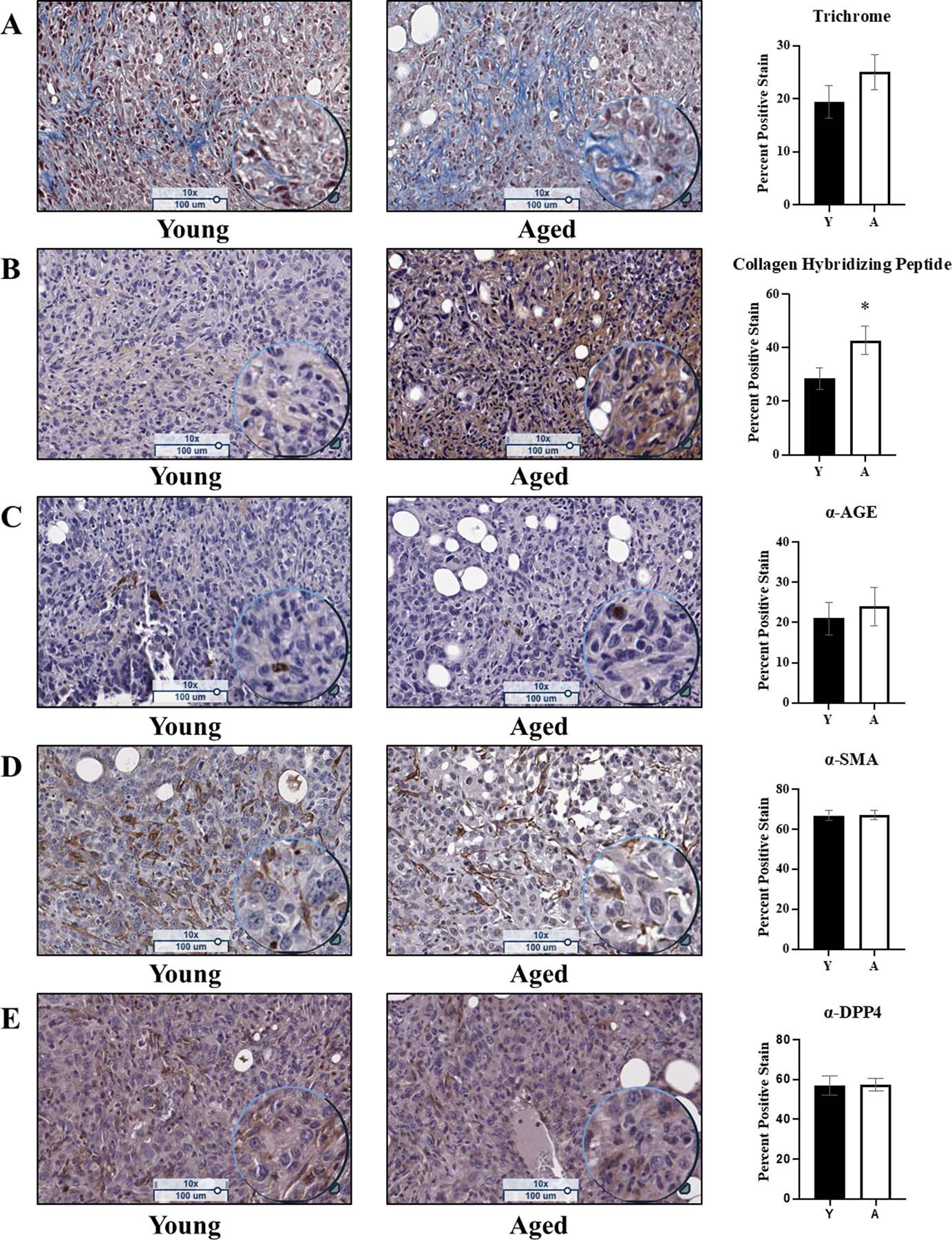
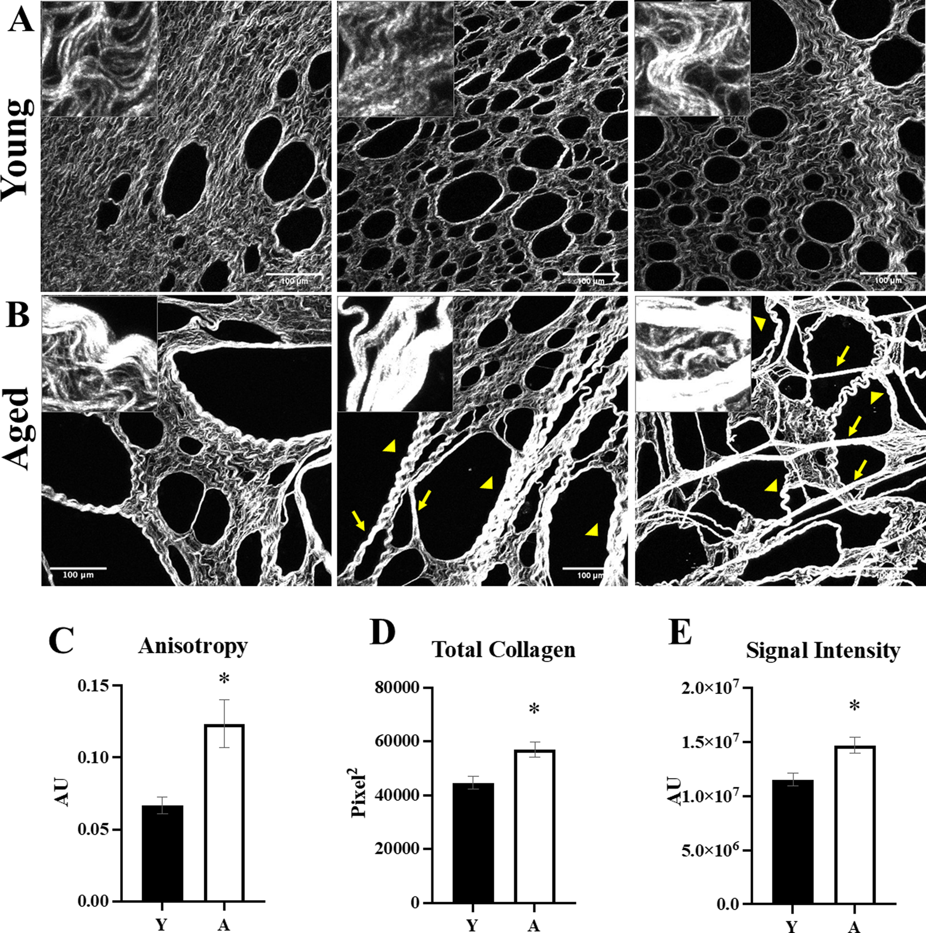
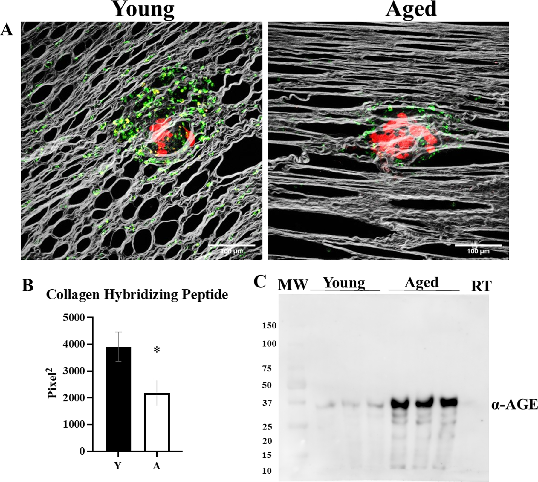
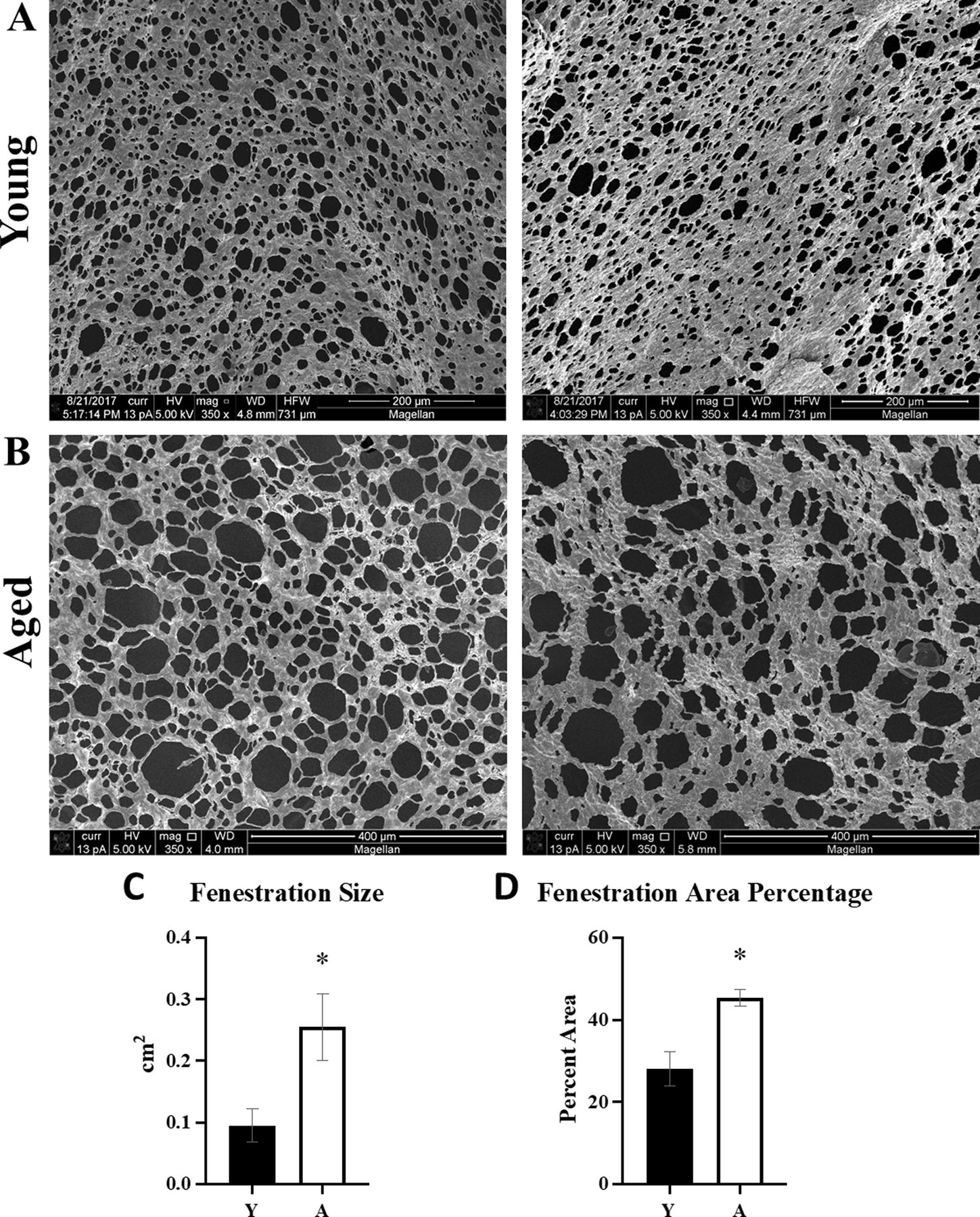
Methods/results: Young (3-6 months) and aged mice (20-23 months) were used to study the role of aging in metastatic success. Intra-peritoneal (IP) injection of ID8Trp53 -/- ovarian cancer cells showed enhanced IP dissemination in aged vs young mice. In vitro assays using purified collagen demonstrated reduced collagenolysis of aged fibers, as visualized using scanning electron microscopy (SEM) and quantified with a hydroxyproline release assay. Omental tumors in young and aged mice showed similar collagen deposition; however enhanced intra-tumoral collagen remodeling was seen in aged mice probed with a biotinylated collagen hybridizing peptide (CHP). In contrast, second harmonic generation (SHG) microscopy showed significant differences in collagen fiber structure and organization in omental tissue and SEM demonstrated enhanced omental fenestration in aged omenta. Combined SHG and Alexa Fluor-CHP microscopy in vivo demonstrated that peri-tumoral collagen was remodeled more extensively in young mice. This collagen population represents truly aged host collagen, in contrast to intra-tumoral collagen that is newly synthesized, likely by cancer associated fibroblasts (CAFs).

Conclusions: Our results demonstrate that tumors in an aged host can grow with minimal collagen remodeling, while tumors in the young host must remodel peri-tumoral collagen to enable effective proliferation, providing a mechanism whereby age-induced ultrastructural changes in collagen and collagen-rich omenta establish a permissive pre-metastatic niche contributing to enhanced OvCa metastatic success in the aged host.

Keywords: aging; collagen; metastasis; omentum; ovarian cancer.


Conflict of interest statement

The authors have stated explicitly that there are no conflicts of interest in connection with this article.

Figures

Figure 1.
Figure 1.. Enhanced growth of OvCa peritoneal metastases in aged mice.
C57Bl/6 mice at 3–6 months of age (Young; Y, n=15) or 20–23 months of age (Aged; A, n=14) were injected IP with RFP-tagged syngeneic ID8Trp53−/− cells (107). Tumors were allowed to grow for 5.5 weeks, then mice were sacrificed and (A) the peritoneal cavity was exposed for in situ fluorescence imaging and (B) omenta were dissected and imaged ex vivo. RFP signal was quantified for (C) abdominal tumor burden (D) and omental tumor burden. (E) Ascites was collected post-sacrifice and the volume was measured. *p<0.005.
Figure 2.
Figure 2.. Analysis of collagen from young vs aged mice.
Collagen was isolated from young (Y) or aged (A) mice. (A,B) Invasion assays. Collagen was used to coat Boyden invasion chambers, followed by addition of the ovarian cancer cell lines (A) OVCAR5 or (B) OVCAR8 (2.5×10⁵) to the wells for 8 or 24 hr, respectively. Invasion was quantified by enumerating cells on the bottom of the filter (n=3, p=0.02 OVCAR5, p=0.22 OVCAR8). (C) Degradation of collagen fibers. Whole tail tendons (n=9) from Y or A mice were incubated with pre-activated MMP-1 (100uM, 16 hr). The supernatant was collected and assayed for hydroxyproline content (p=0.09 as determined by Student’s t-test). Error bars represent standard error of mean. (D) SEM of collagen degradation. Tendons from (C) were collected for scanning electron microscopy (5000x; inset 25,000x). (E) Cleavage products from (C) were electrophoresed on a 9% SDS-PAGE gel and stained with Coomassie to show the ¾ and ¼ fragments of the cleaved collagen. Lanes are: (1) molecular weight markers, (2) 0 μg collagen, (3) 4 μg collagen, (4) 6 μg collagen, (5) 8 μg collagen, (6) 10 μg collagen, (7) 12.5μg collagen, (8) 15 μg collagen, 20μg collagen.
Figure 3.
Figure 3.. Histological and immunohistochemical analysis of omental tumors from young vs aged mice.
Omental tumors from young (n=15) and aged (n=13) C57Bl/6 mice were formalin fixed, paraffin embedded, and sectioned at 5μm width and stained with: (A) Trichrome, a marker for cell nuclei (purple), cell cytoplasm (pink), and collagen (blue), where the blue stain was quantified (p=0.23); (B) Affinity staining with the biotin-conjugated collagen hybridizing peptide, a marker for denatured collagen (brown) and counterstained with hematoxylin (cell nuclei; purple), where the brown signal was quantified (p=0.04); (C) Immunohistochemical analysis of advanced glycation end products (AGEs; brown) and counterstained with hematoxylin (cell nuclei; purple), where the positive brown stain was quantified (p=0.64); (D) Immunohistochemical analysis of smooth muscle actin (SMA; brown), a marker of cancer associated fibroblasts, and counterstained with hematoxylin (cell nuclei; purple), where the positive brown stain was quantified (p=0.89); (E) Immunohistochemical analysis of dipeptidyl-peptidase 4 (DPP4; brown), a marker of activated fibroblasts, and counterstained with hematoxylin (cell nuclei; purple), where the positive brown stain was quantified (p=0.53). Error bars represent standard error of mean and p-values were determined by Student’s t-test.
Figure 4.
Figure 4.. Second harmonic generation (SHG) microscopic analysis of tumor-naïve omenta from young and aged mice.
Omenta (n=5 per cohort) were obtained from (A) young (Y) or (B) aged (A) mice and the collagen was imaged with second harmonic generation (SHG) microscopy (25x; inset 100x). Aged omenta have longer, linear fibers (arrows) and thicker banding of fibers (arrowheads) relative to young omenta. Images were analyzed with ImageJ for (C) anisotropy, a measure of linearity (p=0.01); (D) total collagen (p=0.01); and (E) intensity of the SHG signal (p=0.01). Error bars represent standard error of mean and p-values were determined by Student’s t-test.
Figure 5.
Figure 5.. Visualization of peri-tumoral collagen remodeling in young and aged mice.
C57Bl/6 mice (n=5 per cohort) at 3–6 months of age (Young; Y) or 20–23 months of age (Aged; A) were injected IP with RFP-tagged syngeneic ID8trp53−/− cells (107). Tumors were allowed to grow for 3 weeks, then mice were injected with CHP-B:Streptavidin-Alexa Fluor 647 and sacrificed 3 hr post injection and omenta were imaged with SHG microscopy in conjunction with 2-photon excitation fluorescence microscopy to (A) visualize collagen (grey), tumor cells (red), and biotin conjugate of the collagen hybridizing peptide bound to Alexa Fluor 647 conjugated to streptavidin (pseudocolored green). (B) The images were analyzed for collagen hybridizing peptide signal (p=0.04). (C) Collagen isolated from young and aged mice, as well as commercially available rat tail (RT) collagen, was electrophoresed on a 9% SDS-PAGE gel, transferred to a PVDF membrane, probed with anti-AGE (1:500) and developed with a peroxidase-conjugated secondary antibody (anti-rabbit, 1:4000) and ECL detection. Error bars represent standard error of mean and p-values were determined by Student’s t-test.
Figure 6.
Figure 6.. Analysis of omental fenestration in young vs aged mice.
Tumor-naive omenta (n=5) were obtained from (A) young (Y) or (B) aged (A) FVB/NJ mice and the collagen was imaged with SEM. Images were analyzed with ImageJ for (C) size of the fenestrations in the aged tissue as compared to young (p=0.03), and (D) overall area of the tissue represented by the fenestrations in aged compared to young (p=0.005). Error bars represent standard error of mean and p-values were determined by Student’s t-test.

References

    1. Howlader N et al. , “SEER Cancer Statistics Review, 1975–2014,” Natl. Cancer Inst, vol. Bethesda, MD, Apr. 2017, [Online]. Available: https://seer.cancer.gov/csr/1975_2014/
    1. Yancik R, “Ovarian cancer: Age contrasts in incidence, histology, disease stage at diagnosis, and mortality,” Cancer, vol. 71, no. S2, pp. 517–523, doi: 10.1002/cncr.2820710205. - DOI - PubMed
    1. Loughran EA et al. , “Aging Increases Susceptibility to Ovarian Cancer Metastasis in Murine Allograft Models and Alters Immune Composition of Peritoneal Adipose Tissue,” Neoplasia, vol. 20, no. 6, pp. 621–631, Jun. 2018, doi: 10.1016/j.neo.2018.03.007. - DOI - PMC - PubMed
    1. Cohen CA, Shea AA, Heffron CL, Schmelz EM, and Roberts PC, “The Parity-Associated Microenvironmental Niche in the Omental Fat Band Is Refractory to Ovarian Cancer Metastasis,” Cancer Prev. Res. (Phila. Pa.), vol. 6, no. 11, pp. 1182–1193, Nov. 2013, doi: 10.1158/1940-6207.CAPR-13-0227. - DOI - PMC - PubMed
    1. Lengyel E, “Ovarian Cancer Development and Metastasis,” Am. J. Pathol, vol. 177, no. 3, pp. 1053–1064, Sep. 2010, doi: 10.2353/ajpath.2010.100105. - DOI - PMC - PubMed