. 2022 Sep 15:13:951860.
doi: 10.3389/fphar.2022.951860. eCollection 2022.

Zhuifeng tougu capsules inhibit the TLR4/MyD88/NF-κB signaling pathway and alleviate knee osteoarthritis: In vitro and in vivo experiments

Affiliations

Zhuifeng tougu capsules inhibit the TLR4/MyD88/NF-κB signaling pathway and alleviate knee osteoarthritis: In vitro and in vivo experiments

Xiaotong Xu et al. Front Pharmacol. .

Abstract

Background: Knee osteoarthritis (KOA), a chronic degenerative disease, is mainly characterized by destruction of articular cartilage and inflammatory reactions. At present, there is a lack of economical and effective clinical treatment. Zhuifeng Tougu (ZFTG) capsules have been clinically approved for treatment of OA as they relieve joint pain and inflammatory manifestations. However, the mechanism of ZFTG in KOA remains unknown. Purpose: This study aimed to investigate the effect of ZFTG on the TLR4/MyD88/NF-κB signaling pathway and its therapeutic effect on rabbits with KOA. Study design: In vivo, we established a rabbit KOA model using the modified Videman method. In vitro, we treated chondrocytes with IL-1β to induce a pro-inflammatory phenotype and then intervened with different concentrations of ZFTG. Levels of IL-1β, IL-6, TNF-α, and IFN-γ were assessed with histological observations and ELISA data. The effect of ZFTG on the viability of chondrocytes was detected using a Cell Counting Kit-8 and flow cytometry. The protein and mRNA expressions of TLR2, TLR4, MyD88, and NF-κB were detected using Western blot and RT-qPCR and immunofluorescence observation of NF-κB p65 protein expression, respectively, to investigate the mechanism of ZFTG in inhibiting inflammatory injury of rabbit articular chondrocytes and alleviating cartilage degeneration. Results: The TLR4/MyD88/NF-κB signaling pathway in rabbits with KOA was inhibited, and the levels of IL-1β, IL-6, TNF-α, and IFN-γ in blood and cell were significantly downregulated, consistent with histological results. Both the protein and mRNA expressions of TLR2, TLR4, MyD88, NF-κB, and NF-κB p65 proteins in that nucleus decreased in the ZFTG groups. Moreover, ZFTG promotes the survival of chondrocytes and inhibits the apoptosis of inflammatory chondrocytes. Conclusion: ZFTG alleviates the degeneration of rabbit knee joint cartilage, inhibits the apoptosis of inflammatory chondrocytes, and promotes the survival of chondrocytes. The underlying mechanism may be inhibition of the TLR4/MyD88/NF-kB signaling pathway and secretion of inflammatory factors.

Keywords: NF-κB; TLR4; Zhuifeng tougu capsules; inflammatory factors; innate immune; knee osteoarthritis.


Conflict of interest statement

Author GK was employed by The First Hospital of Hunan University of Chinese Medicine and Hinye Pharmaceutical Co., Ltd. The remaining authors declare that the research was conducted in the absence of any commercial or financial relationships that could be construed as a potential conflict of interest.

Figures

FIGURE 1
FIGURE 1
Structure layout diagram of the automatic production line of ZFTG. Description of reference numeral: 1. a liquid medicine extracting device; 2. double filter; 3. concentration device; 4. ingredient tank; 5. drying device; 6. a crushing device; 7. mixing device; 8. automatic packaging device; 11. a first extraction tank; 12. a second extraction tank; 13. transfer pump; 14. a first liquid storage tank; 15. a second liquid storage tank; 16. oil-water separator; 31. double-effect concentrator; 32. spherical scraper concentrator; 311. a single-effect heating chamber; 312. a primary-effect evaporation chamber; 313. two-effect heating chamber; 314. two-effect evaporation chamber; 51. trough mixer; 52. vacuum drying oven; 53. coating device; 54. vacuum belt drier; 61. mixing hopper; 62. crusher; 63. filtration device; 64. vacuum pump. Patent application number: CN 215133530 U.
FIGURE 2
FIGURE 2
UPLC chromatograms of ZFTG. 6(S): paeoniflorin belongs to Paeonia veitchii Lynch. (Chi Shao, Ranunculaceae) in Northwest China, 4: Gentiana macrophylla Pall. (Qin Jiao, Gentianaceae) in North China, 5: Aconitum kusnezoffii Reichb. (Cao Wu, Ranunculaceae) in North China, 8: Notopterygium incisum Ting. ex H. T. Chang (Qiang Huo, Umbelliferae) in Northwest China, 9: Saposhnikovia divaricata (Turcz.) Schischk. (Fang Feng, Umbelliferae) in Northeast China, 10: Ligusticum chuanxiong Hort. (Chuan Xiong, Umbelliferae) in Sichuan China, 11: Glycyrrhiza uralensis Fisch. (Gan Cao, Fabaceae) in Northwest China.
FIGURE 3
FIGURE 3
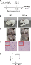
KOA model validation. (A) Experimental design. The KOA model rabbits were treated with the modified Videman modeling. The rabbits were killed 6 weeks after surgery for macroscopic observation and histological analysis (n = 3/group). (B) Outward and (C) anatomical appearance of rabbit knee joint. (D) Representative images and (E) histomorphometric analyses of H&E-stained sections for OARSI score. The images are magnified 100×(inside the red rectangular frame) and 400×. Mann–Whitney test was used for the comparing OARSI scores. The data are presented as the mean ± SD. Specific p-values of comparison are reported.
FIGURE 4
FIGURE 4
Effect of ZFTG on KOA rabbit. (A) Experimental design. The rabbits were killed for H&E analysis at 12 weeks (n = 3/group). (B) Anatomical appearance of rabbit knee joint. (D) Representative images and (C) histomorphometric analyses of H&E-stained sections for OARSI score. The images are magnified 100× (inside the red rectangular frame) and 400×. (E) IL-1β, IL-6, TNF-α, and IFN-γ levels were measured by the ELISA kit according to the manufacturer’s instructions. One-way analysis of variance was used for comparing the OARSI scores. The data are presented as mean ± SD. *p < 0.05, vs. BC group; #p < 0.05, vs. MC group. Specific p-values for each pair of comparisons are reported.
FIGURE 5
FIGURE 5
Immunohistochemistry of IL-1β, IL-6, TNF-α, and IFN-γ. (A) Experimental design. The rabbits were killed for immunohistochemical analysis at 12 weeks (n = 3/group). (B) Representative images and (C) average optical density (AOD) analyses of immunohistochemical sections IL-1β, IL-6, TNF-α, and IFN-γ. The images are magnified 100× (inside the red rectangular frame) and 400×. One-way analysis of variance was used for comparing AOD scores. The data are presented as the mean ± SD. *p < 0.05, vs. BC group; #p < 0.05, vs. MC group. Specific p-values for each pair of comparisons are reported.
FIGURE 6
FIGURE 6
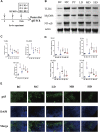
ZFTG inhibits the TLR2-4/MyD88/NF-kB signaling pathway in alleviating knee osteoarthritis in rabbits. (A) Experimental design. The rabbits were killed for Western blot and qRT-PCR analysis at 12 weeks (n = 3/group). (B,C) Western blot analysis of TLR4, MyD88, and NF-kB protein expression with actin serving as a protein loading control. (D) qRT-PCR analysis of TLR2, TLR4, MyD88, and NF-kB mRNA expression. (E) Immunofluorescence observation of NF-κB p65 protein expression. The images are magnified 400×. One-way analysis of variance was used for comparing protein and gene expression. The data are presented as the mean ± SD. *p < 0.05, vs. BC group; #p < 0.05, vs the MC group. Specific p-values for each pair of comparisons are reported.
FIGURE 7
FIGURE 7
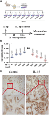
ZFTG enhances the activity of rabbit chondrocytes. (A) Experiment design for the cell. Primary chondrocytes obtained from rabbit knee cartilage (New Zealand rabbits, male, 3-month-old), were treated with ZFTG (0, 50, 100, 200, 400, 800, 1000, 1600, and 2000 ng/μl) to evaluate cell viability. Cells were treated with IL-1β (10 ng/ml) for 24 h s (MIL-1β) to assess inflammation. (B) Cell viability after ZFTG intervention at various concentrations. (C) Expression of collagen II in chondrocytes of the control and IL-1β groups. The images are magnified 100× and 400×. One-way analysis of variance was used for comparing the cell viabilities. The data are presented as the mean ± SD. *p < 0.05, vs. the 400 ng/uL ZFTG group. Specific p-values for each pair of comparisons are reported.
FIGURE 8
FIGURE 8
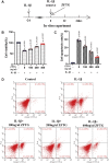
ZFTG improves inflammatory injury of cartilage cells. (A) Experiment design for cells. Primary chondrocytes were stimulated with IL-1β (10 ng/ml) for 24 h s (MIL-1β) before removing IL-1β and adding ZFTG (0, 100, 200, and 400 ng/μl) treatment to evaluate cell viability and apoptosis rate. Cells were harvested 24 h after ZFTG treatment. (B) Cell viability after ZFTG intervention at various concentrations based on the control group (100%). (C) Cell apoptosis rate after ZFTG intervention at various concentrations. (D) Apoptosis of chondrocytes among five groups by flow cytometry. One-way analysis of variance was used for comparing the cell viabilities and apoptosis rates. The data are presented as the mean ± SD. *p < 0.05, vs. the control group; #p < 0.05, vs. IL-1β group. Specific p-values for each pair of comparison are reported. The control group: ZFTG/IL-1β; the IL-1β group: ZFTG/IL-1β+.
FIGURE 9
FIGURE 9
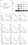
ZFTG inhibits the TLR2/TLR4/MyD88 signaling pathway in alleviating inflammatory damage of chondrocytes. (A) Experiment design for cells. Primary chondrocytes were stimulated with IL-1β (10 ng/ml) for 24 h s (MIL-1β) before removing IL-1β and adding ZFTG (0, 100, 200, and 400 ng/μl) treatment. (B,D) Western blot analysis of TLR2, TLR4, and MyD88 protein expression with actin serving as a protein loading control. (C) IL-1β, IL-6, and TNF-α levels were measured by the ELISA kit according to the manufacturer’s instructions. (E) qRT-PCR analysis of TLR2, TLR4, and MyD88 mRNA expression. One-way analysis of variance was used for comparing the protein and gene expression. The data are presented as the mean ± SD. *p < 0.05, vs. BC group; #p < 0.05, vs. MC group. Specific p-values for each pair of comparisons are reported.

Similar articles

Cited by

References

    1. Ahmad M., Hachemi Y., Paxian K., Mengele F., Koenen M., Tuckermann J. (2019). A jack of all trades: Impact of glucocorticoids on cellular cross-talk in osteoimmunology. Front. Immunol. 10, 2460. 10.3389/fimmu.2019.02460 - DOI - PMC - PubMed
    1. Barreto G., Senturk B., Colombo L., Bruck O., Neidenbach P., Salzmann G., et al. (2020). Lumican is upregulated in osteoarthritis and contributes to TLR4-induced pro-inflammatory activation of cartilage degradation and macrophage polarization. Osteoarthr. Cartil. 28 (1), 92–101. 10.1016/j.joca.2019.10.011 - DOI - PubMed
    1. Bisson D. G., Sheng K., Kocabas S., Krock E., Teles A., Saran N., et al. (2020). Toll-like receptor involvement in adolescent scoliotic facet joint degeneration. J. Cell. Mol. Med. 24 (19), 11355–11365. 10.1111/jcmm.15733 - DOI - PMC - PubMed
    1. Blasioli D. J., Kaplan D. L. (2014). The roles of catabolic factors in the development of osteoarthritis. Tissue Eng. Part B Rev. 20 (4), 355–363. 10.1089/ten.TEB.2013.0377 - DOI - PMC - PubMed
    1. Bonizzi G., Karin M. (2004). The two NF-kappaB activation pathways and their role in innate and adaptive immunity. Trends Immunol. 25 (6), 280–288. 10.1016/j.it.2004.03.008 - DOI - PubMed

LinkOut - more resources