. 2022 Aug 20;13(4):28-39.
eCollection 2022.

Ligand recognition by peptidoglycan recognition protein-S (PGRP-S): structure of the complex of camel PGRP-S with heptanoic acid at 2.15 Å resolution

Affiliations

Ligand recognition by peptidoglycan recognition protein-S (PGRP-S): structure of the complex of camel PGRP-S with heptanoic acid at 2.15 Å resolution

Ankit Maurya et al. Int J Biochem Mol Biol. .

Abstract

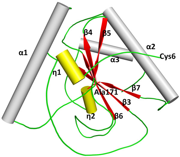
Peptidoglycan recognition proteins (PGRPs) are important components of the innate immune system which provide the first line of defense against invading microbes. There are four members in the family of PGRPs in animals of which PGRP-S is a common domain. It is responsible for the binding to microbial cell wall molecules. In order to understand the mode of binding of PGRP-S to the components of the bacterial cell wall, the structure of the complex of camel PGRP-S (CPGRP-S) with heptanoic acid has been determined at 2.15 Å resolution. The structure determination showed the presence of four crystallographically independent protein molecules which are designated as A, B, C, and D. These four protein molecules associate in the form of two homodimers which are represented as A-B and C-D dimers. The association between molecules A and B gives rise to a shallow cleft on the surface at one end of the dimeric interface. One molecule of heptanoic acid is observed at this binding site in the A-B dimer. The association of C and D molecules results in the formation of a long zig-zag tunnel along with the C-D interface. In the cleft at the C-D interface, three molecules of hydrogen peroxide along with other non-water solvent molecules have been observed. The analysis of the several complexes of CPGRP-S with fatty acids and non-fatty acids such as peptidoglycan, lipopolysaccharide, and lipoteichoic acid shows that the fatty acids bind at the A-B site while non-fatty acids interact through C-D interface.

Keywords: PGRP-S; Peptidoglycan recognition protein; dimers; ligand binding site; structures.


Conflict of interest statement

None.

Figures

Figure 1
Figure 1
Showing the amino acid sequence comparison of PGRP-S from camel, human, C-terminal PGN domain of human, common lancelet, Drosophila, bumblebees, and tobacco hornworm. The segments involved in the intermolecular arrangement between molecules A and B are indicated in red while those responsible for the formation of C-D dimer are shown in green. The critical residues for the formation of A-B dimer are Ser8 and Asn126 (shown in purple) while those in the formation of C-D dimer are Pro96 and Pro151 (shown in blue).
Figure 2
Figure 2
Electron density maps of various ligands with CPGRP-S. (i) The initial (Fo-Fc) map showing electron densities at 3.0 σ cut off for one heptanoic acid, nine hydrogen peroxide, one 2-methyl-2,4-pentanediol, four ethylene glycol, and one carbonate and one sodium ions and (ii) the final (2Fo-Fc) map showing electron densities at 1.0 σ cut off for the above entities.
Figure 3
Figure 3
Structure of CPGRP-Swith four molecules, A, B, C, and D in the asymmetric unit. The bound heptanoic acid at the ligand-binding site in the A-B dimer and several solvent molecules other than the water molecules are indicated in the cleft of the C-D dimer. The numbering scheme of the heptanoic acid is shown at the bottom of the figure.
Figure 4
Figure 4
The secondary structure elements in CPGRP-S are shown. Five β-strands including β3, β4, β5, β6, and β7 are shown in red where β3, β4, β6, and β7 are in the parallel arrangement while β5 is oriented in an antiparallel sense. Three α-helices, α1, α2, and α3 are shown in grey. There are two short helices, η1 and η2 in yellow.
Figure 5
Figure 5
Showing (i) A-B and (ii) C-D dimers with shaded ligand binding sites and (iii) showing the polymeric arrangement of A-B and C-D interfaces.
Figure 6
Figure 6
Showing grasp view of the region at the A-B interface with a bound heptanoic acid in the ball and stick model.
Figure 7
Figure 7
Cα-superimpositions of A-B dimer of CPGRP-S (cyan) on the Cα traces of two crystallographic independent molecules observed in (i) Drosophila (PDB ID, 1XSR, grey) and (ii) bumblebee (PDB ID, 5ZX4, orange).
Figure 8
Figure 8
Superimpositions of Cα traces of (i) A-B dimer of CPGRP-S from the present complex on the Cα traces of the A-B dimer from the complexes of CPGRP-S with bound ligands at the A-B site and (ii) C-D dimer of CPGRP-S from the present complex on the Cα traces of the C-D dimer from the other complexes of CPGRP-S with bound ligands at the C-D site.
Figure 9
Figure 9
Cα-superimpositions of C-D dimer of CPGRP-S (cyan) on the Cα traces of two crystallographic independent molecules observed in (i) Drosophila (PDB ID, 1XSR, grey) and (ii) bumblebee (PDB ID, 5ZX4, orange).

References

    1. Guan R, Mariuzza RA. Peptidoglycan recognition proteins of the innate immune system. Trends Microbiol. 2007;15:127–134. - PubMed
    1. Wang Z, Zhou W, Huang B, Gao M, Li Q, Tao Y, Wang Z. Molecular and functional characterization of peptidoglycan recognition proteins of PGRP-A and of PGRP-B in ostriniafurnacalis (Lepidoptera: Crambidae) Insects. 2022;13:417. - PMC - PubMed
    1. Dziarski R. Peptidoglycan recognition proteins (PGRPs) Mol Immunol. 2004;40:877–886. - PubMed
    1. Hou J, Gan Z, Chen SN, Nie P. Molecular and functional characterization of a short-type peptidoglycan recognition protein, PGRP-S in the amphibian Xenopus laevis. Dev Comp Immunol. 2019;98:13–19. - PubMed
    1. Huang L, Chen SN, Gan Z, Nie P. Molecular and functional identification of a short-type peptidoglycan recognition protein, PGRP-S, in the Chinese soft-shelled turtle Pelodiscus sinensis. Dev Comp Immunol. 2021;117:103965. - PubMed

LinkOut - more resources