

Unusual Presentation of Wünderlich Syndrome
- PMID: 36189085
- PMCID: PMC9477127
- DOI: 10.31486/toj.21.0120
Unusual Presentation of Wünderlich Syndrome
Abstract
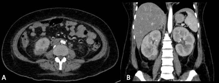
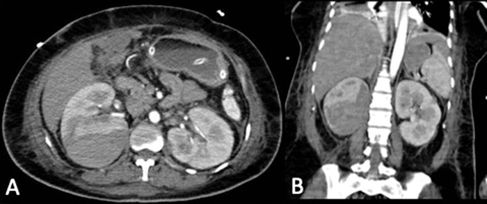
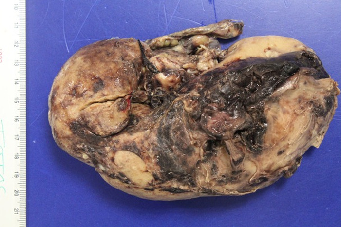
Background: Wünderlich syndrome is a rare but important condition because it involves a sudden blood collection in the renal fossa that can cause hemodynamic instability. Case Report: A 38-year-old female with a history of type 2 diabetes mellitus and hypertension with poor adherence to treatment presented to the emergency department with abdominal pain of 2 weeks' duration accompanied by irritative lower urinary symptoms. Abdominal computed tomography (CT) scan showed bilateral pyelonephritis and an abscess in the lower pole of the right kidney. A second CT scan, performed because of the patient's abrupt decrease in hemoglobin and hematocrit, showed active bleeding secondary to the infectious process in the right kidney. The patient was hemodynamically unstable, so a nephrectomy was performed. Conclusion: Wünderlich syndrome is a spontaneous renal hemorrhage, in most cases attributed to a tumorous etiology and rarely of infectious origin. The clinical picture is varied but can present with the Lenk triad of acute onset flank pain, flank mass, and hypovolemic shock. It is diagnosed principally via an imaging study such as abdominal CT scan. Treatment is conservative in principle, but urgent surgical intervention is sometimes necessary depending on the clinical situation of the patient.
Keywords: Hemorrhage; kidney; nephrectomy; pyelonephritis.
©2022 by the author(s); Creative Commons Attribution License (CC BY).
Figures

References
-
- Sales R, Villa V, Caballé J, et al. . Síndrome de Wünderlich. Hemorragia renal espontánea. Cir Esp. 2000;68(5):493-495.
-
- Bonet T. Sepulchretum Sive Anatomia Practica, Ex Cadaveribus Morbo Denatis. Vol 3. Cramer & Perachon; 1679.
Publication types
LinkOut - more resources
Full Text Sources