. 2022 Sep 9:13:987151.
doi: 10.3389/fimmu.2022.987151. eCollection 2022.

Role of N- Glycosylation in FcγRIIIa interaction with IgG

Affiliations

Role of N- Glycosylation in FcγRIIIa interaction with IgG

Julie Van Coillie et al. Front Immunol. .

Abstract

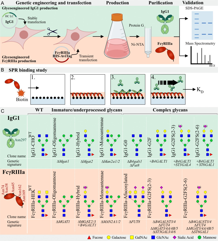
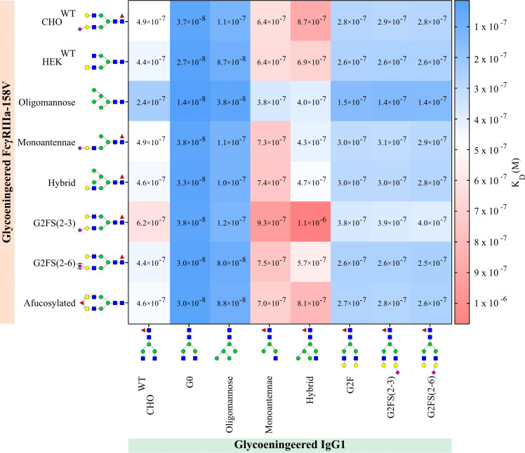
Immunoglobulins G (IgG) and their Fc gamma receptors (FcγRs) play important roles in our immune system. The conserved N-glycan in the Fc region of IgG1 impacts interaction of IgG with FcγRs and the resulting effector functions, which has led to the design of antibody therapeutics with greatly improved antibody-dependent cell cytotoxicity (ADCC) activities. Studies have suggested that also N-glycosylation of the FcγRIII affects receptor interactions with IgG, but detailed studies of the interaction of IgG1 and FcγRIIIa with distinct N-glycans have been hindered by the natural heterogeneity in N-glycosylation. In this study, we employed comprehensive genetic engineering of the N-glycosylation capacities in mammalian cell lines to express IgG1 and FcγRIIIa with different N-glycan structures to more generally explore the role of N-glycosylation in IgG1:FcγRIIIa binding interactions. We included FcγRIIIa variants of both the 158F and 158V allotypes and investigated the key N-glycan features that affected binding affinity. Our study confirms that afucosylated IgG1 has the highest binding affinity to oligomannose FcγRIIIa, a glycan structure commonly found on Asn162 on FcγRIIIa expressed by NK cells but not monocytes or recombinantly expressed FcγRIIIa.

Keywords: CD16a; Fc gamma receptors; IgG; N-glycosylation; glycoengineering; glycosyltransferases; mAbs; surface plasmon resonance.


Conflict of interest statement

The University of Copenhagen has filed a patent application for the cell-based display platform. GlycoDisplay Aps, Copenhagen, Denmark, has obtained a license in the field of the patent application. Authors YN, ZY, and HC are co-founders of GlycoDisplay Aps and hold ownerships in the company. The remaining authors declare that the research was conducted in the absence of any commercial or financial relationships that could be construed as a potential conflict of interest.

Figures

Figure 1
Figure 1
Graphic depiction of glycoengineered IgG1 and FcγRIIIa production, validation, and binding studies. (A) Top: Production of CHOWT stably expressing IgG1. In this parental clone, KO and KI of glycosyltransferases resulted in a library of genetically glycoengineered CHO clones stably producing IgG1. Expressed IgG1 was purified by protein G and validated by SDS-PAGE and Mass Spectrometry (MS). Bottom, HEK293 cells were genetically engineered to display distinct N-glycan structures and this library of genetically engineered HEK clones were transiently transfected with soluble HIS- and AviTag-tagged FcγRIIIa-158F/V. Produced FcγRIIIa was purified by Ni-NTA columns and subjected to SDS-PAGE and MS for validation. (B) Schematic depiction of the IBIS MX96 SPR setup. 1) All glycoengineered FcγRIIIa glycoforms were enzymatically biotinylated by BirA and spotted at four different concentrations on a streptavidin-coated chip. 2) Glycoengineered IgG1 was injected at eight different dilutions, 3) allowing for binding affinity measurements of each antibody to all glycoengineered FcγRIIIa’s simultaneously. 4) Regeneration after every sample was carried out after which the next IgG1 glycoform was injected. (C) Glycoengineered IgG1 and FcγRIIIa-158F/V expressing cell lines with clone name, gene editing background and expected N-glycan signature based on gene editing signature and literature. IgG1 has one N-glycan site (Asn297) per Fc domain and FcγRIIIa has 5 N-glycan sites (Asn38, Asn45, Asn74, Asn162, and Asn169). Designations for monosaccharides are according to the Consortium for Functional Glycomics (CFG) (51).
Figure 2
Figure 2
SDS-PAGE of purified recombinant IgG1 and FcγRIIIa-158F/V. Reducing SDS-PAGE of 1 μg purified (A) glycoengineered IgG1 produced in CHO cells with the heavy chain and light chain at ~50 and 30 kDa, respectively. (B) HEK293-expressed His- and AviTag-tagged FcγRIIIa-158F (left lane) and -158V (right lane) migrating as a broad band with a molecular weight ranging from 40 to 50 kDa. Designations for monosaccharides according to the CFG are indicated (51).
Figure 3
Figure 3
Site-specific N-glycan profiling of Asn162 FcγRIIIa-158F/V by LC-MS/MS. FcγRIIIa-158F and -158V expressed in (A) CHO and (B–H) HEK293 cells were digested by a combination of chymotrypsin and Glu-C and subjected to LC-MS/MS. Site-specific N-glycan profiling of Asn162 was carried out and the top 90% of the structures are represented relative to the most abundant structure (A.U. = 1) for FcγRIIIa-158F (dark grey, left) and -158V (light grey, right) with H: hexose, N: N-acetylhexosamine, F: fucose; S: Sialic acid. Proposed glycan structures are based on gene editing signatures and literature. Structures are depicted following the CFG notation (51).
Figure 4
Figure 4
IgG1 and FcγRIIIa-158V binding. Surface plasmon resonance dissociation constant (KD) was determined from SPR analysis after biotinylated glycoengineered FcγRIIIa-158V was spotted at 4 concentrations and bound to glycoengineered IgG1 at 8 times dilution series. Calculation of the dissociation constant was performed by equilibrium fitting to Rmax= 500. Mean data is reported of three independent experiments for each IgG1:FcγRIIIa-158V pair.

References

    1. Baković M, Selman H, Hoffmann H, Rudan I, Campbell H, Deelder A, et al. . High-throughput IgG fc n-glycosylation profiling by mass spectrometry of glycopeptides. J Proteome Res (2013) 12:821–31. doi: 10.1021/PR300887Z - DOI - PubMed
    1. Stadlmann J, Pabst M, Kolarich D, Kunert R, Altmann F. Analysis of immunoglobulin glycosylation by LC-ESI-MS of glycopeptides and oligosaccharides. Proteomics (2008) 8:2858–71. doi: 10.1002/pmic.200700968 - DOI - PubMed
    1. Barb AW, Brady EK, Prestegard JH. Branch specific sialylation of IgG-fc glycans by ST6Gal-I. Biochemistry (2009) 48:9705. doi: 10.1021/BI901430H - DOI - PMC - PubMed
    1. Guddat LW, Herron JN, Edmundson AB. Three-dimensional structure of a human immunoglobulin with a hinge deletion. Proc Natl Acad Sci U.S.A. (1993) 90:4271. doi: 10.1073/PNAS.90.9.4271 - DOI - PMC - PubMed
    1. Barb AW, Prestegard JH. NMR analysis demonstrates immunoglobulin G n-glycans are accessible and dynamic. Nat Chem Biol (2011) 7:147–53. doi: 10.1038/NCHEMBIO.511 - DOI - PMC - PubMed

Publication types

LinkOut - more resources