. 2022 Sep 16:12:990402.
doi: 10.3389/fcimb.2022.990402. eCollection 2022.

Psychological stress creates an immune suppressive environment in the lung that increases susceptibility of aged mice to Mycobacterium tuberculosis infection

Affiliations

Psychological stress creates an immune suppressive environment in the lung that increases susceptibility of aged mice to Mycobacterium tuberculosis infection

William P Lafuse et al. Front Cell Infect Microbiol. .

Abstract

Age is a major risk factor for chronic infections, including tuberculosis (TB). Elderly TB patients also suffer from elevated levels of psychological stress. It is not clear how psychological stress impacts immune response to Mycobacterium tuberculosis (M.tb). In this study, we used social disruption stress (SDR) to investigate effects of psychological stress in young and old mice. Unexpectedly, we found that SDR suppresses lung inflammation in old mice as evidenced by lower pro-inflammatory cytokine levels in bronchial lavage fluid and decreased cytokine mRNA expression by alveolar macrophages. To investigate effects of stress on M.tb infection, mice were subjected to SDR and then infected with M.tb. As previously reported, old mice were better at controlling infection at 30 days than young mice. This control was transient as CFUs at 60 days were higher in old control mice compared to young mice. Consistently, SDR significantly increased M.tb growth at 60 days in old mice compared to young mice. In addition, SDR in old mice resulted in accumulation of IL-10 mRNA and decreased IFN-γ mRNA at 60 days. Also, confocal microscopy of lung sections from old SDR mice showed increased number of CD4 T cells which express LAG3 and CD49b, markers of IL-10 secreting regulatory T cells. Further, we also demonstrated that CD4 T cells from old SDR mice express IL-10. Thus, we conclude that psychological stress in old mice prior to infection, increases differentiation of IL-10 secreting T cells, which over time results in loss of control of the infection.

Keywords: IL10; Mycobacterium tuberculosis; aging; granuloma; inflammaging; social stress.


Conflict of interest statement

The authors declare that the research was conducted in the absence of any commercial or financial relationships that could be construed as a potential conflict of interest.

Figures
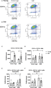
Figure 1
Figure 1
Flow cytometry analysis of myeloid cells in BAL fluid from Home Cage and SDR mice. CD11c/CD11b flow plots of BAL cells from young (A) and old (B) mice. CD11c+ CD11b- AMs are in quadrant 1. CD11c+ CD11b+ AMs are in quadrant 2. Monocytes and neutrophils present in the CD11c- CD11b+ cells (quadrant 4) were detected using antibodies to Ly6G and Ly6C. Cell numbers of each population present in BAL fluid were determined. (C) CD11c+ CD11b- AM population (D) CD11c+ CD11b+ AM population (E) Neutrophils (F) Monocytes. Each symbol represents cells present in individual mice. Statistical analysis was performed by one-way ANOVA with Tukey multiple-comparisons post hoc test. N=4-5 mice/group. Data are shown as mean ± SE. *p <0.05, *** p < 0.001. ns, non significant.
Figure 2
Figure 2
SDR in old mice suppresses cytokine levels in BAL fluid and basal mRNA levels in alveolar macrophages. BAL fluid and alveolar macrophages were isolated from individual young (3 months) and old (18 months) Home Cage mice and mice subjected to 6 cycles of SDR. (A–C) Cytokine levels in BAL fluid determined by ELISA. Protein levels in BAL fluid were determined by Bio-RAD protein assay (D). (E–G) Basal cytokine mRNA expression in AMs. RNA was isolated from individual mice without culture and mRNA levels determined by qRT-PCR. Expression levels were normalized to β-actin mRNA. Protein levels in BAL fluid were determined by Bio-RAD protein assay. (H, I) Corticosterone and Norepinephrine levels in BAL fluid determined by ELISA. Each symbol represents individual mice. N=9-15 mice/group. Data are shown as mean ± SE. Statistical analysis was performed by one-way ANOVA with Tukey multiple-comparisons post hoc test. ns, non significant. * p < 0.05, ** p < 0.01, ****p < 0.0001.
Figure 3
Figure 3
SDR suppresses TLR2 induced pro-inflammatory cytokine mRNA levels in AMs from old mice. AMs were pooled from six old Home Cage and six SDR mice were cultured at 37° for 2 hrs. Nonadherent cells were removed by washing and adherent cells stimulated with TLR2 ligand Pam3CSK4 (100 ng/ml) for 6 hrs. RNA was isolated and cytokine mRNA levels determined by qRT-PCR. (A-D) RNA levels are expressed relative to β-actin mRNA levels. Results are representative of two independent experiments with 3 wells/treatment. Data are shown as mean ± SE. Statistical analysis was performed by one-way ANOVA with Tukey multiple-comparisons post hoc test. * p < 0.05.
Figure 4
Figure 4
Old mice initially control M.tb infection better than young mice but eventually lose control of M.tb infection, which is further exacerbated by SDR. Young and old Home Cage mice subjected to six cycles of SDR were infected with low dose aerosol of M. tuberculosis. At 30 days (A) and 60 days (B), the left lung lobes were homogenized and M. tuberculosis CFUs enumerated by plating on 7H11 duplicate plates. Results are representative of two independent experiments with 3 mice/group per time point. Data are shown as mean ± SE. Statistical analysis was performed by one-way ANOVA with Tukey multiple-comparisons post hoc test. **p < 0.01, *** p < 0.001, ****p < 0.0001.
Figure 5
Figure 5
mRNA levels of cytokines genes in the lungs of mice infected with M.tb. Young and old Home Cage mice and mice subjected to six cycles of SDR were infected with low dose aerosol of M. tuberculosis for 30 and 60 days. RNA was isolated from a right lung lobe and mRNA levels determined by qRT-PCR (A-I). RNA levels are expressed relative to β-actin. Results are pooled from two independent experiments. Each symbol represents individual mice. Data are shown as mean ± SE. Statistical analysis was performed by one-way ANOVA with Tukey multiple-comparisons post hoc test. * p< 0.05, ** p < 0.01, ***p < 0.001. ns, non significant.
Figure 6
Figure 6
mRNA levels of macrophage and T cell genes in the lungs of mice infected with M.tb. Young and old Home Cage mice and mice subjected to six cycles of SDR were infected with low dose aerosol of M. tuberculosis for 30 and 60 days. RNA was isolated from a right lung lobe and mRNA levels (A–F) determined by qRT-PCR. RNA levels are expressed relative to β-actin. Each symbol represents individual mice. Data are shown as mean ± SE. Statistical analysis was performed by one-way ANOVA with Tukey multiple-comparisons post hoc test. Results represent 3 mice/group per time point. Data are shown as mean ± SE. * p< 0.05, ** p < 0.01, ***p < 0.001, ****p < 0.0001. ns, non significant.
Figure 7
Figure 7
SDR in old mice up-regulate expression of LAG3 and CD49b in CD4+T cells (A) Representative images of LAG3 expression in CD4+ T cells present in regions of pneumonia (B) Representative images of CD49b in CD4+ T cells. (C) Granulomatous tissue in old SDR mice contain more CD4 T cells. CD4 T cells were counted in regions of granulomatous tissue. Each symbol represents a region of granulomatous tissue. N=15 regions/group. Mean fluorescence intensity of LAG3 (D) and CD49b (E) expression in CD4 T cells was determined using Image (J) N= 90 cells/group. Statistical analysis was performed by one-way ANOVA with Tukey multiple-comparisons post hoc test. ****p < 0.0001. ns, non significant.
Figure 8
Figure 8
SDR in old mice results in production of IL-10 by CD4+ T cells in the lungs of M.tb infected mice. Lung tissues sections from mice infected with M.tb for 60 days were examined by confocal immunofluorescence microscopy for expression of IL-10 (red fluorescence) in CD4+ T cells (green fluorescence) present in areas of pneumonia. (A) Representative images of IL-10 expression in CD4+ T cells. (B) Mean fluorescence of IL-10 expression in CD4 T cells was determined using Image (J) N= 90 cells/group. Statistical analysis was performed by one-way ANOVA with Tukey multiple-comparisons post hoc test. ****p < 0.0001.

Similar articles

Cited by

References

    1. Abdalla A. E., Li Q., Xie L., Xie J. (2015). Biology of IL-27 and its role in the host immunity against mycobacterium tuberculosis. Int. J. Biol. Sci. 11, 168–175. doi: 10.7150/ijbs.10464 - DOI - PMC - PubMed
    1. Agac D., Estrada L. D., Maples R., Hooper L. V., Farrar J. D. (2018). The beta2-adrenergic receptor controls inflammation by driving rapid IL-10 secretion. Brain Behavior Immun. 74, 176–185. doi: 10.1016/j.bbi.2018.09.004 - DOI - PMC - PubMed
    1. Allen R. G., Lafuse W. P., Galley J. D., Ali M. M., Ahmer B. M., Bailey M. T. (2012). The intestinal microbiota are necessary for stressor-induced enhancement of splenic macrophage microbicidal activity. Brain Behavior Immun. 26, 371–382. doi: 10.1016/j.bbi.2011.11.002 - DOI - PMC - PubMed
    1. Allen R. G., Lafuse W. P., Powell N. D., Webster Marketon J. I., Stiner-Jones L. M., Sheridan J. F., et al. . (2012). Stressor-induced increase in microbicidal activity of splenic macrophages is dependent upon peroxynitrite production. Infection Immun. 80, 3429–3437. doi: 10.1128/IAI.00714-12 - DOI - PMC - PubMed
    1. Avitsur R., Stark J. L., Sheridan J. F. (2001). Social stress induces glucocorticoid resistance in subordinate animals. Hormones Behav. 39, 247–257. doi: 10.1006/hbeh.2001.1653 - DOI - PubMed

Publication types