Review
. 2022 Nov 1;323(5):C1444-C1474.
doi: 10.1152/ajpcell.00246.2022. Epub 2022 Oct 3.

Different modalities of host cell death and their impact on Mycobacterium tuberculosis infection

Affiliations
Review

Different modalities of host cell death and their impact on Mycobacterium tuberculosis infection

Annuurun Nisa et al. Am J Physiol Cell Physiol. .

Abstract

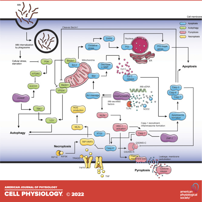
Mycobacterium tuberculosis (Mtb) is the pathogen that causes tuberculosis (TB), a leading infectious disease of humans worldwide. One of the main histopathological hallmarks of TB is the formation of granulomas comprised of elaborately organized aggregates of immune cells containing the pathogen. Dissemination of Mtb from infected cells in the granulomas due to host and mycobacterial factors induces multiple cell death modalities in infected cells. Based on molecular mechanism, morphological characteristics, and signal dependency, there are two main categories of cell death: programmed and nonprogrammed. Programmed cell death (PCD), such as apoptosis and autophagy, is associated with a protective response to Mtb by keeping the bacteria encased within dead macrophages that can be readily phagocytosed by arriving in uninfected or neighboring cells. In contrast, non-PCD necrotic cell death favors the pathogen, resulting in bacterial release into the extracellular environment. Multiple types of cell death in the PCD category, including pyroptosis, necroptosis, ferroptosis, ETosis, parthanatos, and PANoptosis, may be involved in Mtb infection. Since PCD pathways are essential for host immunity to Mtb, therapeutic compounds targeting cell death signaling pathways have been experimentally tested for TB treatment. This review summarizes different modalities of Mtb-mediated host cell deaths, the molecular mechanisms underpinning host cell death during Mtb infection, and its potential implications for host immunity. In addition, targeting host cell death pathways as potential therapeutic and preventive approaches against Mtb infection is also discussed.

Keywords: cell death signaling; granuloma; host-directed therapy; immune cells; inflammation.


Conflict of interest statement

No conflicts of interest, financial or otherwise, are declared by the authors.

Figures

None
Graphical abstract
Figure 1.
Figure 1.
Intrinsic and extrinsic host apoptosis pathways in Mycobacterium tuberculosis infection. Apoptosis is induced by the host’s innate immune response against infection by either intrinsic or extrinsic pathways. A range of mycobacterial proteins inhibits host-induced apoptosis during successful infection. Pathogen-associated molecular patterns (PAMPs) are recognized by host signaling proteins that drive the cell toward apoptosis. Specific bacterial factors cause mitochondrial membrane depolarization and lead to the release of mitochondrial factors that trigger apoptosis by the intrinsic pathway. Various stimuli can induce intrinsic apoptosis by shifting the equilibrium of prosurvival B-cell lymphoma-2 (Bcl-2) and Bcl‐2 homology 3 (BH3) proteins. Extrinsic apoptosis can be induced by the binding of a select group of tumor necrosis factor (TNF) family ligands to their receptors leading to death-inducing signaling complex (DISC) formation by recruitment of adapter Fas‐associated protein with death domain (FADD)/TNFRSF1A‐associated via death domain (TRADD) and caspase-8. Caspase-8 auto-processes itself (cCasp8—cleaved/activated caspase-8) and can directly activate caspase-3 or cleave Bid to generate tBid and triggers intrinsic apoptosis. Apaf-1, apoptotic peptidase activating factor 1; Bax, B-cell leukemia/lymphoma-2 associated-X, Bak, B-cell leukemia/lymphoma-2 antagonist/killer; BID, BH3 interacting domain; Bok, B-cell leukemia/lymphoma-2 associated related ovarian killer; CAD, intracellular caspase-activated deoxyribonuclease; CARD, caspase recruitment domain; Casp, caspase; CytC, cytochrome C; FAD, flavin adenine dinucleotide; FASL, FAS ligand; iFAS, FS-7-associated surface antigen; LAM, lipoarabinomannan; SMAC, second mitochondria-derived activator of caspases; tBID, truncated BID; TNFα, tumor necrosis factor-alpha; TNFR, tumor necrosis factor-alpha receptor; XIAP, X-linked inhibitor of apoptosis protein. Figure created with images from smart.servier.com.
Figure. 2.
Figure. 2.
Mycobacterium tuberculosis (Mtb) modulation of host autophagy signaling. Multiple surface receptors and intracellular stress signals, including Mtb factors, modulate autophagy signaling. Microbial surface pattern receptors such as Toll-like receptors (TLRs), RIG-I like receptors (RLRs), and NOD-like receptors (NLRs) recognize mycobacterial secreted factors, which further activate p38 mitogen-activated protein kinases (MAPK) and receptor-interacting serine/threonine-protein kinase 1 (RIP1)/extracellular signal-regulated kinases (ERK) that phosphorylate phosphoinositide 3-kinases (PI3K) and signal Beclin-1 phosphorylation to trigger autophagy. Autophagy culminates in the fusion of lysosomes with autophagosomes. Early secreted antigenic target of 6 kDa (ESAT-6), lipoarabinomannan (LAM), enhanced intracellular survival protein 2 (Eis), serine/threonine-protein phosphatase 2 A activator (PtpA), protein kinase G (PknG), early secretion system-1 (Esx-1), LAM/mannose-capped lipoarabinomannan (ManLAM) inhibit autophagy. P in the blue circle represents forward signaling by phosphorylation, while P in the red circle represents inhibition of phosphorylation and, thereby, inhibition of downstream signaling. Following phagocytosis, Mtb resides in the phagosome and blocks phagosome maturation. Mtb secretes Esx-1, promoting phagosome damages that trigger ubiquitination, recruitment of autophagic adaptors and mycobacterial capture via stimulator of interferon genes (STING). NOD, nucleotide binding and oligomerization domain; RIG-I, retinoic acid-inducible gene-I. Figure created with images from smart.servier.com.
Figure 3.
Figure 3.
Cross talk of mycobacterial factors during different forms of host cell death. Various cell death modalities may result from either mycobacteria-induced events or host-induced innate defense mechanisms. The dynamics of these modalities determine the outcome of Mycobacterium tuberculosis (Mtb) infection. Several host factors are involved in multiple cell death pathways, and Mtb factors can inhibit host defense mechanisms at various levels. Arrows indicate activation, deactivation, and forward signaling, the color of the signaling components defines the particular pathway as indicated. The pathways are simplified to accommodate the cross connections showing the mycobacterial factors that increase the complexity of multiple cell death networks in eliminating mycobacteria. ASC-1, alanine/serine/cysteine transporter-1; ATG3, autophagy related 3; Bcl-2, B-cell leukemia/lymphoma 2; Bid, BH3 interacting domain; Casp, caspase; cGAMP, cyclic guanosine monophosphate/adenosine monophosphate; CytC, cytochrome C; DAMPs, death associated molecular patterns; DAPK, death associated protein kinase; eDNA, extracellular DNA; ER, endoplasmic reticulum; GSDMD, Gasdermin D; LC3, light chain 3; MLKL, mixed lineage kinase domain-like pseudokinase; MMP, matrix metalloproteinase; mTORC, mammalian target of rapamycin-C; NLR, NOD-like receptor; NOD, nucleotide binding oligomerization domain; p53, protein 53; PAMPs, pathogen associated molecular patterns; PI3K, phosphatidylinositol 3-kinase; RIPK, receptor interacting serine/threonine kinase; ROS, reactive oxygen species; tBid, truncated BH3 interacting domain; TBK, Tank-binding kinase; TLR, Toll-like receptor; TNF, tumor necrosis factor-alpha; TNFR, tumor necrosis factor-alpha receptor; TRADD, TNF receptor superfamily member 1 A associated via death domain; ULK, Unc-51 like autophagy activating kinase; VPS34, vacuolar protein sorting 34; ZMP-1, zinc metalloproteinase-1. Figure created with images from smart.servier.com.

References

    1. World Health Organization. Global Tuberculosis Report 2021. Geneva: WHO, 2021.
    1. Fukunaga R, Glaziou P, Harris JB, Date A, Floyd K, Kasaeva T. Epidemiology of tuberculosis and progress toward meeting global targets—worldwide, 2019. MMWR Morb Mortal Wkly Rep 70: 427–430, 2021. doi: 10.15585/mmwr.mm7012a4. - DOI - PMC - PubMed
    1. Green DR, Llambi F. Cell death signaling. Cold Spring Harb Perspect Biol 7: a006080, 2015. doi: 10.1101/cshperspect.a006080. - DOI - PMC - PubMed
    1. Galluzzi L, Vitale I, Aaronson SA, Abrams JM, Adam D, Agostinis P, et al. Molecular mechanisms of cell death: recommendations of the Nomenclature Committee on Cell Death 2018. Cell Death Differ 25: 486–541, 2018. doi: 10.1038/s41418-017-0012-4. - DOI - PMC - PubMed
    1. Tang D, Kang R, Berghe TV, Vandenabeele P, Kroemer G. The molecular machinery of regulated cell death. Cell Res 29: 347–364, 2019. doi: 10.1038/s41422-019-0164-5. - DOI - PMC - PubMed

Publication types