

Evaluation of visual field changes with retinal nerve fiber layer thickness in primary congenital glaucoma
- PMID: 36190046
- PMCID: PMC9789852
- DOI: 10.4103/ijo.IJO_396_22
Evaluation of visual field changes with retinal nerve fiber layer thickness in primary congenital glaucoma
Abstract
Purpose: To evaluate visual field changes in primary congenital glaucoma (PCG) with retinal nerve fiber layer thickness on optical coherence tomography.
Methods: In this cross-sectional, observational study, consecutive PCG children who underwent combined trabeculotomy with trabeculectomy and on regular follow-up were enrolled. All patients were aged over four years and co-operative for RNFL OCT and visual field examination. Perimetry was done on Humphrey visual field (HVF) analyzer using 30-2 and 10-2 SITA standard algorithms as appropriate. If a reliable automated perimetry was not feasible, kinetic perimetry was done. The following were noted at baseline and every follow-up: age, sex, visual acuity, intraocular pressure (IOP), cup-disc ratio (CDR), corneal diameters, refraction, any topical antiglaucoma medications, surgeries underwent, age at surgery and duration between surgery and final examination.
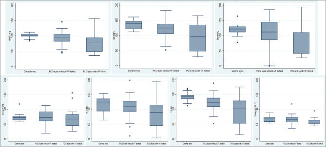
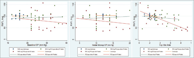
Results: Forty-eight eyes of 34 children operated for PCG and 19 eyes of 17 controls were analyzed. A statistically significant thinner average RNFL thickness of 87.2 ± 28 μm was noted in PCG eyes as compared to controls with 100.6 ± 7.2 μm (P = 0.04). The mean cup-disc area ratio on OCT in PCG eyes was 0.43 ± 0.2 (0.02-0.93) and in control eyes was 0.23 ± 0.07 (0.1-0.4) (P < 0.001). On RNFL OCT, there was significant focal RNFL loss in temporal superior (P = 0.003), nasal inferior (P = 0.037) and temporal inferior (P < 0.001) quadrants compared to controls. Among PCG eyes, 20/48 eyes (41.7%), had definitive, reproducible glaucomatous VF defects. Mean baseline IOP in PCG eyes with VF defect was 28.7 ± 5.7 mmHg and in eyes with normal VF was 24.6 ± 5.9 mmHg (P = 0.03). On univariate regression analysis, higher baseline IOP was significantly associated with both RNFL loss (odds ratio (OR): -2.17) and VF defects (OR: 3.35). Fluctuation in follow-up IOP (OR: 3.33) was also significantly associated with the presence of VF defects. On multivariable regression analysis maximum, IOP was significantly associated with RNFL loss and VF defects.
Conclusion: Peripapillary RNFL thickness could be used to identify PCG eyes having visual field loss and possibly poor visual function from PCG eyes without visual field defects. Baseline and follow-up IOP, significantly correlated with RNFL thickness in PCG eyes.
Keywords: RNFL OCT in Primary congenital glaucoma; RNFL thickness in primary congenital glaucoma; visual field loss in PCG.
Conflict of interest statement
None
Figures
Similar articles
-
Association between Intraocular Pressure and Rates of Retinal Nerve Fiber Layer Loss Measured by Optical Coherence Tomography.Ophthalmology. 2016 Oct;123(10):2058-65. doi: 10.1016/j.ophtha.2016.07.006. Epub 2016 Aug 20. Ophthalmology. 2016. PMID: 27554036 Free PMC article. Clinical Trial.
-
Retinal Nerve Fiber Layer Optical Texture Analysis: Detecting Axonal Fiber Bundle Defects in Patients with Ocular Hypertension.Ophthalmology. 2023 Oct;130(10):1080-1089. doi: 10.1016/j.ophtha.2023.06.004. Epub 2023 Jun 12. Ophthalmology. 2023. PMID: 37315588
-
Severity of visual field defects in primary congenital glaucoma and their risk factors.Graefes Arch Clin Exp Ophthalmol. 2020 Jul;258(7):1483-1491. doi: 10.1007/s00417-020-04677-w. Epub 2020 Apr 15. Graefes Arch Clin Exp Ophthalmol. 2020. PMID: 32296990
-
Advancing glaucoma detection with convolutional neural networks: a paradigm shift in ophthalmology.Rom J Ophthalmol. 2023 Jul-Sep;67(3):222-237. doi: 10.22336/rjo.2023.39. Rom J Ophthalmol. 2023. PMID: 37876506 Free PMC article. Review.
-
Consensus on Outcome Measures for Glaucoma Effectiveness Trials: Results From a Delphi and Nominal Group Technique Approaches.J Glaucoma. 2016 Jun;25(6):539-46. doi: 10.1097/IJG.0000000000000301. J Glaucoma. 2016. PMID: 26091178
Cited by
-
Potential Involvements of Cilia-Centrosomal Genes in Primary Congenital Glaucoma.Int J Mol Sci. 2024 Sep 18;25(18):10028. doi: 10.3390/ijms251810028. Int J Mol Sci. 2024. PMID: 39337513 Free PMC article.
-
Evaluating retinal thickness classification in children: A comparison between pediatric and adult optical coherence tomography databases.PLoS One. 2024 Dec 30;19(12):e0314395. doi: 10.1371/journal.pone.0314395. eCollection 2024. PLoS One. 2024. PMID: 39775315 Free PMC article.
References
-
- Moore DB, Tomkins O, Ben-Zion I. A review of primary congenital glaucoma in the developing world. Surv Ophthalmol. 2013;58:278–85. - PubMed
-
- Dandona L, Dandona R, Naduvilath TJ, McCarty CA, Nanda A, Srinivas M, et al. Is current eye-care-policy focus almost exclusively on cataract adequate to deal with blindness in India? Lancet Lond Engl. 1998;351:1312–6. - PubMed
-
- Zagora SL, Funnell CL, Martin FJ, Smith JEH, Hing S, Billson FA, et al. Primary congenital glaucoma outcomes:Lessons From 23 Years of Follow-up. Am J Ophthalmol. 2015;159:788–96.e2. - PubMed
Publication types
MeSH terms
LinkOut - more resources
Full Text Sources
Medical
