

Peripapillary and fovea avascular zone optical coherence tomography angiography parameters in exfoliation glaucoma versus primary open-angle glaucoma versus healthy eyes
- PMID: 36190047
- PMCID: PMC9789868
- DOI: 10.4103/ijo.IJO_84_22
Peripapillary and fovea avascular zone optical coherence tomography angiography parameters in exfoliation glaucoma versus primary open-angle glaucoma versus healthy eyes
Abstract
Purpose: To examine the differences in the peripapillary vascular parameters and foveal-avascular-zone (FAZ) vascularity parameters between primary open-angle-glaucoma (POAG) patients versus exfoliation-glaucoma (XFG) patients versus healthy subjects.
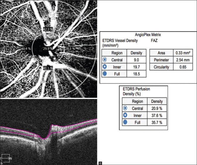
Methods: This is cross-sectional study and a comparative clinical study. POAG and XFG patients and healthy subjects underwent a comprehensive ophthalmic examination, including visual field optical coherence tomography (OCT) and OCT angiography (OCTA) of the optic disc and FAZ. Differences in peripapillary vessel density (VD), perfusion density (PD), and FAZ area and circularity were examined between all groups, as well as correlations between clinical parameters and vascularity parameters for each glaucoma group.
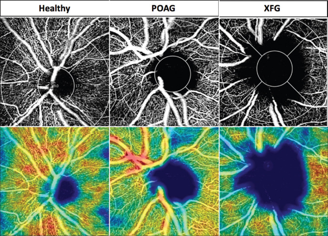
Results: A total of 109 subjects (one eye for each patient) were analyzed, including 45 with POAG, 30 with XFG, and 34 controls. The average peripapillary VDs were the lowest among the XFG patients and the highest among the controls (P < 0.05, ANOVA). The average peripapillary PD of the central ring was the lowest in the XFG group and the highest in the control group (P = 0.02, ANOVA). A significant negative correlation was found between the average peripapillary VDs and PDs of the inner ring and full ring and disease severity of the POAG patients. There was a significant positive correlation between the average peripapillary PDs of the central rings and full ring and the central macular thickness of the XFG patients (P < 0.01 and P < 0.04, respectively, Pearson correlation).
Conclusion: The peripapillary vascular parameters of the POAG and XFG patients were lower compared to those of normal participants. A correlation between clinical characteristics of POAG and XFG patients and PD was found. This may hint to a vascular mechanism in glaucoma either primary or secondary to intra-ocular pressure/OAG damage.
Keywords: OCTA; POAG; PXF.
Conflict of interest statement
None
Figures
Similar articles
-
Macula Vessel Density and Foveal Avascular Zone Parameters in Exfoliation Glaucoma Compared to Primary Open-Angle Glaucoma.Invest Ophthalmol Vis Sci. 2019 Mar 1;60(4):1244-1253. doi: 10.1167/iovs.18-25986. Invest Ophthalmol Vis Sci. 2019. PMID: 30924849 Free PMC article.
-
Comparison of vessel density in macular and peripapillary regions between primary open-angle glaucoma and pseudoexfoliation glaucoma using OCTA.Int Ophthalmol. 2021 Jan;41(1):173-184. doi: 10.1007/s10792-020-01564-5. Epub 2020 Aug 26. Int Ophthalmol. 2021. PMID: 32851558
-
Comparison of microvascular parameters and diagnostic ability of optical coherence tomography angiography between eyes with primary angle closure glaucoma and primary open angle glaucoma.Photodiagnosis Photodyn Ther. 2022 Dec;40:103114. doi: 10.1016/j.pdpdt.2022.103114. Epub 2022 Sep 10. Photodiagnosis Photodyn Ther. 2022. PMID: 36096437
-
A comprehensive update on the use of optical coherence tomography angiography in glaucoma.Int Ophthalmol. 2023 May;43(5):1785-1802. doi: 10.1007/s10792-022-02574-1. Epub 2022 Dec 6. Int Ophthalmol. 2023. PMID: 36472722 Review.
-
The Role of Vessel Density as Measured by Optical Coherence Tomography Angiography in the Evaluation of Pseudoexfoliative Glaucoma: A Review of the Literature.Ophthalmol Ther. 2022 Apr;11(2):533-545. doi: 10.1007/s40123-022-00483-1. Epub 2022 Feb 24. Ophthalmol Ther. 2022. PMID: 35211880 Free PMC article. Review.
Cited by
-
Early changes of ganglion cell-inner plexiform layer thickness and macular microvasculature in Posner-Schlossman syndrome: a binocular control study by OCTA.Front Med (Lausanne). 2023 Jul 24;10:1169504. doi: 10.3389/fmed.2023.1169504. eCollection 2023. Front Med (Lausanne). 2023. PMID: 37554506 Free PMC article.
References
-
- Weinreb RN, Khaw PT. Primary open-angle glaucoma. Lancet (London England) 2004;363:1711–20. - PubMed
-
- Tanna AP. Growing evidence of the importance of the macula in glaucoma. JAMA Ophthalmol. 2017;135:747–8. - PubMed
-
- Leske MC, Heijl A, Hyman L, Bengtsson B, Dong L, Yang Z, et al. Predictors of long-term progression in the early manifest glaucoma trial. Ophthalmology. 2007;114:1965–72. - PubMed
MeSH terms
LinkOut - more resources
Full Text Sources
Miscellaneous
