. 2022 Oct 26;13(5):e0180422.
doi: 10.1128/mbio.01804-22. Epub 2022 Oct 3.

Structural and Mechanistic Bases of Viral Resistance to HIV-1 Capsid Inhibitor Lenacapavir

Affiliations

Structural and Mechanistic Bases of Viral Resistance to HIV-1 Capsid Inhibitor Lenacapavir

Stephanie M Bester et al. mBio. .

Abstract

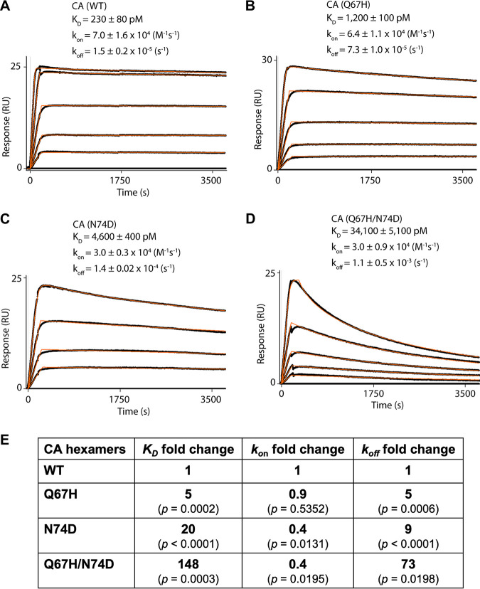
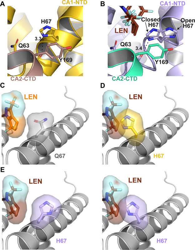
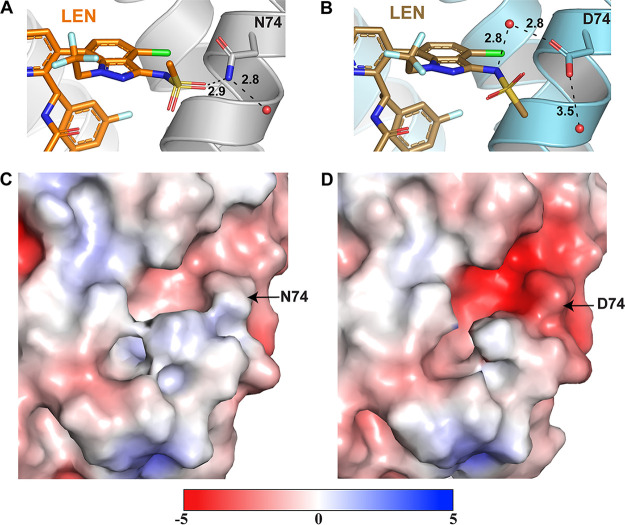
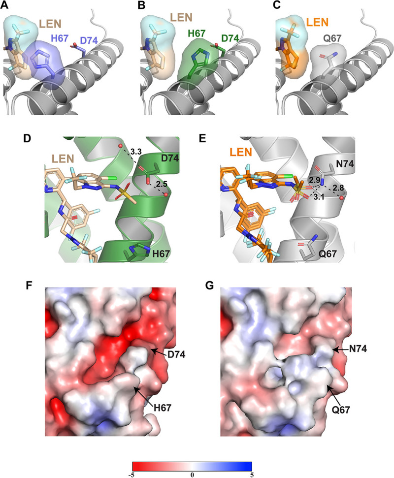
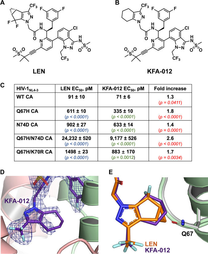
Lenacapavir (LEN) is a long-acting, highly potent HIV-1 capsid (CA) inhibitor. The evolution of viral variants under the genetic pressure of LEN identified Q67H, N74D, and Q67H/N74D CA substitutions as the main resistance associated mutations (RAMs). Here, we determined high-resolution structures of CA hexamers containing these RAMs in the absence and presence of LEN. Our findings reveal that the Q67H change induces a conformational switch, which adversely affects the inhibitor binding. In the unliganded protein, the His67 side chain adopts the closed conformation by projecting into the inhibitor binding pocket and thereby creating steric hindrance with respect to LEN. Upon the inhibitor binding, the His67 side chain repositions to the open conformation that closely resembles the Gln67 side chain in the WT protein. We propose that the switch from the closed conformation to the open conformation, which is needed to accommodate LEN, accounts for the reduced inhibitor potency with respect to the Q67H CA variant. The N74D CA change results in the loss of a direct hydrogen bond and in induced electrostatic repulsions between CA and LEN. The double Q67H/N74D substitutions exhibited cumulative effects of respective single amino acid changes. An examination of LEN binding kinetics to CA hexamers revealed that Q67H and N74D CA changes adversely influenced the inhibitor binding affinity (KD) by primarily affecting the dissociation rate constant (koff). We used these structural and mechanistic findings to rationally modify LEN. The resulting analog exhibited increased potency against the Q67H/N74D viral variant. Thus, our studies provide a means for the development of second-generation inhibitors with enhanced barriers to resistance. IMPORTANCE LEN is an investigational long-acting agent for future HIV-1 treatment regimens. While ongoing clinical trials have highlighted a largely beneficial profile of LEN for the treatment of HIV-1 infected people with limited therapy options, one notable shortcoming is a relatively low barrier of viral resistance to the inhibitor. Cell culture-based viral breakthrough assays identified N74D, Q67H, and N74D/Q67H capsid changes as the main resistance associated mutations (RAMs). N74D and Q67H capsid substitutions have also emerged in clinical trials in some patients who received subcutaneous LEN. Understanding the structural basis behind viral resistance to LEN is expected to aid in the rational development of improved inhibitors with enhanced barriers to resistance. Here, we report high resolution structures of the main drug resistant capsid variants, which provide mechanistic insight into the viral resistance to LEN. We used these findings to develop an improved inhibitor, which exhibited enhanced activity against the viral Q67H/N74D capsid phenotype compared with that of parental LEN.

Keywords: HIV-1; antiretroviral agents; capsid; drug resistance mechanisms; structure.


Conflict of interest statement

The authors declare no conflict of interest.

Figures

FIG 1
FIG 1
LEN binding to cross-linked HIV-1 CA hexamers. (A to D). Representative binding sensorgrams illustrating interactions between LEN with wild-type (WT) and mutant cross-linked HIV-1 CA hexamers evaluated by SPR detection. The binding data (black lines) were fit (orange lines) to a simple kinetic model with a mass transport added as needed. The mean ± standard deviation (SD) equilibrium dissociation constant (KD), association rate constant (kon), and dissociation rate constant (koff) values were determined from three independent experiments with comparable results. The LEN concentrations are 0.156, 0.312, 0.625, 1.25, and 2.5 μM in (A); 0.078, 0.156, 0.312, 0.625, and 1.25 μM in (B); 0.156, 0.312, 0.625, and 1.25 μM in (C); and 0.078, 0.156, 0.312, 0.625, and 2.5 μM in (D). (E) Fold change of the KD, kon, and koff values for the mutant CA variants compared to WT CA. P values indicate differences compared to CA(WT) hexamers.
FIG 2
FIG 2
Structural basis for the LEN resistance of the CA(Q67H) hexamer. (A) X-ray crystal structure of the prestabilized CA(Q67H) hexamer (gold/dirty violet; PDB: 7RAR) with interactions between His67 and Tyr169 and Gln63 displayed. Hydrogen bonding interactions are denoted by black dashed lines, and distances are indicated. (B) X-ray crystal structure of prestabilized CA(Q67H) + LEN (lilac/seafoam + chocolate; PDB: 7RHN) displaying two conformations of His67, open and closed, and interactions between His67 and Tyr169 and Gln63. (C) Crystal structure of CA + LEN (light gray + orange; PDB: 6VKV) with surface renderings of LEN and Gln67 to demonstrate how they interact. (D) Overlay of CA(Q67H) (gold) and CA(Q67H) + LEN (not shown + chocolate) crystal structures with surface renderings of LEN and His67 to demonstrate how they would potentially interact. The darkened/shadowed surface rendering denotes an overlap in the space occupied by His67 and LEN. Cartoon regions are shown in light gray to highlight the substituted residue and LEN. (E) Crystal structure of CA(Q67H) + LEN (lilac + chocolate) with surface renderings of LEN and both conformations of His67 to demonstrate how they would potentially interact. The darkened/shadowed surface rendering denotes an overlap in the space occupied by His67 and LEN.
FIG 3
FIG 3
Structural basis for the LEN resistance of the CA(N74D) hexamer. (A) X-ray crystal structure of CA + LEN (light gray + orange; PDB: 6VKV) denoting the hydrogen bonding interaction between LEN and residue N74. Hydrogen bonding interactions are denoted by black dashed lines, and respective distances are indicated. (B) X-ray crystal structure of prestabilized CA(N74D) + LEN (steel blue + sand; PDB: 7RJ4) highlighting the hydrogen bonding interaction between LEN and residue Asp74 via a water molecule. (C and D) Surface rendering of the CA for CA(WT) (PDB: 3H47) and CA(N74D) (PDB: 7RMM), respectively, with the surface potential ranging from −5 to +5. Negatively charged regions are shown in red, and positively charged regions are shown in blue. Potentials were generated via the PDB2PQR server, and the surface was rendered using the adaptive Poisson-Boltzmann solver (APBS).
FIG 4
FIG 4
Structural basis for the LEN resistance of the CA(Q67H/N74D) hexamer. (A) X-ray crystal structure of the apo CA(Q67H/N74D) hexamer (slate purple; PDB: 7RHM) superimposed onto the CA(Q67H/N74D) + LEN (not shown + wheat) crystal structure with surface renderings of LEN and His67 to demonstrate how they would potentially interact. The darkened/shadowed surface rendering denotes an overlap in the space occupied by His67 and LEN. Cartoon regions are shown in light gray to highlight the substituted residue and LEN. (B) X-ray crystal structure of CA(Q67H/N74D) + LEN (forest green + wheat) with surface renderings of LEN and His67 to demonstrate their interaction. (C) Crystal structure of CA + LEN (light gray + orange; PDB: 6VKV) with surface renderings of LEN and Gln67 to demonstrate how they interact. (D) X-ray crystal structure of the prestabilized CA(Q67H/N74D) hexamer + LEN (forest green + wheat; PDB: 7RJ2), illustrating the lack of interactions between LEN and residue Asp74. (E) X-ray crystal structure of CA + LEN (light gray + orange; PDB: 6VKV) denoting the hydrogen bonding interaction between LEN and residue N74. Hydrogen bonding interactions are denoted by black dashed lines, and respective distances are indicated. (F and G) Surface renderings of the CA1-NTD for CA(Q67H/N74D) (PDB: 7RHM) and CA(WT) (PDB: 3H47), respectively, with the surface potential ranging from −5 to +5. Negatively charged regions are shown in red, and positively charged regions are shown in blue. Potentials were generated via the PDB2PQR server, and the surface was rendered using the adaptive Poisson-Boltzmann solver (APBS).
FIG 5
FIG 5
Comparison of the antiviral activities of LEN and KFA-012 against WT and CA mutants. (A) Chemical structure of LEN. (B) Chemical structure of the modified LEN-like compound KFA-012. (C) Early stage antiviral activities of LEN and KFA-012 were measured against pseudotyped HIV-1NL4-3_RFP in MT-4 cells. P values in blue and green indicate differences compared to WT for LEN and KFA-012, respectively; P values in red show the statistical significance for the fold increases of the KFA-012 potency versus LEN potency with respect to the indicated CA variants. (D) X-ray crystal structure of the cross-linked CA(WT) hexamer (light green/peach) in complex with KFA-012 (dark purple) with the 2Fo-Fc density scaled to 1σ (blue mesh; PDB: 7RMJ). (E) Superimposition of cross-linked CA(WT) hexamer in complex with KFA-012 (PDB: 7RMJ; light green + dark purple) and LEN (PDB 6VKV; gray + orange).

References

    1. Link JO, Rhee MS, Tse WC, Zheng J, Somoza JR, Rowe W, Begley R, Chiu A, Mulato A, Hansen D, Singer E, Tsai LK, Bam RA, Chou CH, Canales E, Brizgys G, Zhang JR, Li J, Graupe M, Morganelli P, Liu Q, Wu Q, Halcomb RL, Saito RD, Schroeder SD, Lazerwith SE, Bondy S, Jin D, Hung M, Novikov N, Liu X, Villasenor AG, Cannizzaro CE, Hu EY, Anderson RL, Appleby TC, Lu B, Mwangi J, Liclican A, Niedziela-Majka A, Papalia GA, Wong MH, Leavitt SA, Xu Y, Koditek D, Stepan GJ, Yu H, Pagratis N, Clancy S, Ahmadyar S, et al. . 2020. Clinical targeting of HIV capsid protein with a long-acting small molecule. Nature 584:614–618. doi:10.1038/s41586-020-2443-1. - DOI - PMC - PubMed
    1. Yant SR, Mulato A, Hansen D, Tse WC, Niedziela-Majka A, Zhang JR, Stepan GJ, Jin D, Wong MH, Perreira JM, Singer E, Papalia GA, Hu EY, Zheng J, Lu B, Schroeder SD, Chou K, Ahmadyar S, Liclican A, Yu H, Novikov N, Paoli E, Gonik D, Ram RR, Hung M, McDougall WM, Brass AL, Sundquist WI, Cihlar T, Link JO. 2019. A highly potent long-acting small-molecule HIV-1 capsid inhibitor with efficacy in a humanized mouse model. Nat Med 25:1377–1384. doi:10.1038/s41591-019-0560-x. - DOI - PMC - PubMed
    1. Selyutina A, Hu P, Miller S, Simons LM, Yu HJ, Hultquist JF, Lee K, KewalRamani VN, Diaz-Griffero F. 2022. GS-CA1 and lenacapavir stabilize the HIV-1 core and modulate the core interaction with cellular factors. iScience 25:103593. doi:10.1016/j.isci.2021.103593. - DOI - PMC - PubMed
    1. Bester SM, Wei G, Zhao H, Adu-Ampratwum D, Iqbal N, Courouble VV, Francis AC, Annamalai AS, Singh PK, Shkriabai N, Van Blerkom P, Morrison J, Poeschla EM, Engelman AN, Melikyan GB, Griffin PR, Fuchs JR, Asturias FJ, Kvaratskhelia M. 2020. Structural and mechanistic bases for a potent HIV-1 capsid inhibitor. Science 370:360–364. doi:10.1126/science.abb4808. - DOI - PMC - PubMed
    1. Sowd GA, Shi J, Aiken C. 2021. HIV-1 CA inhibitors are antagonized by inositol phosphate stabilization of the viral capsid in cells. J Virol 95:e0144521. doi:10.1128/JVI.01445-21. - DOI - PMC - PubMed

Publication types