. 2022 Oct 26;7(5):e0033322.
doi: 10.1128/msphere.00333-22. Epub 2022 Oct 3.

ALIBY: ALFA Nanobody-Based Toolkit for Imaging and Biochemistry in Yeast

Affiliations

ALIBY: ALFA Nanobody-Based Toolkit for Imaging and Biochemistry in Yeast

Dipayan Akhuli et al. mSphere. .

Abstract

Specialized epitope tags continue to be integral components of various biochemical and cell biological applications such as fluorescence microscopy, immunoblotting, immunoprecipitation, and protein purification. However, until recently, no single tag could offer this complete set of functionalities on its own. Here, we present a plasmid-based toolkit named ALIBY (ALFA toolkit for imaging and biochemistry in yeast) that provides a universal workflow to adopt the versatile ALFA tag/NbALFA system within the well-established model organism Saccharomyces cerevisiae. The kit comprises tagging plasmids for labeling a protein of interest with the ALFA tag and detection plasmids encoding fluorescent-protein-tagged NbALFA for live-cell imaging purposes. We demonstrate the suitability of ALIBY for visualizing the spatiotemporal localization of yeast proteins (i.e., the cytoskeleton, nucleus, centrosome, mitochondria, vacuole, endoplasmic reticulum, exocyst, and divisome) in live cells. Our approach has yielded an excellent signal-to-noise ratio without off-target effects or any effect on cell growth. In summary, our yeast-specific toolkit aims to simplify and further advance the live-cell imaging of differentially abundant yeast proteins while also being suitable for biochemical applications. IMPORTANCE In yeast research, conventional fluorescent protein tags and small epitope tags are widely used to study the spatiotemporal dynamics and activity of proteins. Although proven to be efficient, these tags lack the versatility for use across different cell biological and biochemical studies of a given protein of interest. Therefore, there is an urgent need for a unified platform for visualization and biochemical and functional analyses of proteins of interest in yeast. Here, we have engineered ALIBY, a plasmid-based toolkit that expands the benefits of the recently developed ALFA tag/NbALFA system to studies in the well-established model organism Saccharomyces cerevisiae. We demonstrate that ALIBY provides a simple and versatile strain construction workflow for long-duration live-cell imaging and biochemical applications in yeast.

Keywords: Saccharomyces cerevisiae; biochemistry; cell biology; cell division; fluorescence; fluorescent image analysis; live-cell imaging; mitochondria; nanobody; vacuoles.


Conflict of interest statement

The authors declare no conflict of interest.

Figures

FIG 1
FIG 1
Design of detection and tagging plasmids comprising the toolkit and the proposed workflow. (a) Schematic map of a representative tagging plasmid (top) and its naming scheme (bottom). The ALFA tag amino acid sequence is (P)SRLEEELRRRLTE. The number in the plasmid name code corresponds to the yeast selectable marker, as indicated. (b) Schematic map of a representative pRS-based detection plasmid (top) and its naming scheme (bottom). 40aaL, 40-amino-acid linker; mNG, mNeonGreen. AmpR encodes a β-lactamase responsible for conferring resistance to ampicillin, used as a bacterial selectable marker. The black dashed line marks the linearization site in the yeast selectable marker gene. In the plasmid name code, the first digit corresponds to the promoter, while the second digit corresponds to the yeast selectable marker, as indicated. (c) Schematic of the recommended workflow for strain construction. The S2 primer overhang sequence is 5′-ATCGATGAATTCGAGCTCG-3′; the S3 primer overhang sequence is 5′-CGTACGCTGCAGGTCGAC-3′. Starting from a particular strain of choice (middle left), the PCR fragment generated from the tagging plasmid using S2/S3 primers (top left) can be transformed to generate the tagged strain (middle center). The linearized detection plasmid (top right) containing a promoter and the selectable marker of choice can now be integrated into the tagged strain to obtain the nanobody-containing tagged strain (middle right), which can be used for live-cell imaging (bottom right). The colored blocks in the top row correspond to those already labeled in panels a and b. The color-code for schematics in the bottom row is as follows: pink, POI; purple, ALFA tag; orange, NbALFA; fluorescent green, dye; brown, 40-amino-acid linker; green, mNeonGreen. FACS, fluorescence-activated cell sorting.
FIG 2
FIG 2
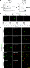
Characterization of plasmids, immunoprecipitation, and live-cell imaging of various proteins. (a) Immunoprecipitation (IP) of Gin4ALFA, Bud4ALFA, and Exo84ALFA. The wild-type control strain lacks the ALFA tag. Total cell extracts (TCE) of each strain were incubated with ALFA SelectorST resin and washed; 1/50 of the input fraction and the entire eluate fraction were resolved by SDS-PAGE and analyzed by immunoblotting using anti-ALFA sdAb and antiactin (n = 3) (* indicates an unspecific band). (b) Comparison of promoters for optimizing NbALFA expression by laser point scanning confocal microscopy. (Top row) ALFA-tagged Shs1 in ESM356 cells carrying pRS305-promoter-NbALFA-L-mNG-termCYC1. Shown from left to right are PTEF1, PGPD, PADH1, and PCYC1. Images were captured at 1% laser excitation power at 488 nm. (Bottom row) Intensity profiles for cells with Shs1ALFA expressing NbALFA-L-mNG under the control of different promoters in the same order as the one described above. Bar, 5 μm. a.u., arbitrary units. (c) Images of ALFA-tagged proteins. From left to right (in each row) are the ALFA-tagged POI along with NbALFA-L-mNG under the control of PTEF1 (green), the mCherry-tagged marker protein (red), and the merged image. First row, Shs1, a terminal septin, with Cdc3-mCherry as a bud neck marker; second row, Spc42, a spindle pole body component, with mCherry-Tub1 as a microtubule marker; third row, Emc1, an ER membrane protein, with Elo3mCherry as an ER marker; fourth row, Om45, a mitochondrial outer membrane protein, with Cox4-mCherry as a mitochondrial marker; fifth row, Exo84, an exocyst complex component, with Cdc3-mCherry as a bud neck marker. All of the strains were grown in synthetic complete (SC) medium at 30°C. All of the images were maximum-intensity projected and deconvolved for representation. Bar, 5 μm.
FIG 3
FIG 3
Time-lapse imaging and quantification of protein kinetics. The time interval between successive images shown in each montage is denoted as Δt. (a) Bud4-GFP (top) and Bud4ALFA (bottom) (Δt = 8 min). (b) Bni5-GFP (top) and Bni5ALFA (bottom) (Δt = 8 min). (c) Shs1-GFP (top) and Shs1ALFA (bottom) (Δt = 3 min). (d) Signal-to-noise ratio comparison between C-terminal ALFA tag and GFP fusions of Bni5, Bud4, and Shs1 (n = 3 replicates; the number of cells quantified was 36 to 42 per strain). (e) Comparison of the kinetics of Bud4-GFP and Bud4ALFA (n = 3 replicates; the number of cells quantified was 49 to 53 per strain). (f) Comparison of the kinetics of Bni5-GFP and Bni5ALFA (n = 3 replicates; the number of cells quantified was 49 to 53 per strain). (g) Comparison of the kinetics of Shs1-GFP and Shs1ALFA (n = 3 replicates; the number of cells quantified was 49 to 53 per strain). In addition to the ALFA-tagged proteins, the above-mentioned ALFA tag-containing strains also expressed NbALFA-L-mNG under the control of PTEF1. All of the strains were grown in SC medium at 30°C. Green, ALFA-tagged proteins bound to NbALFA-L-mNG and GFP-tagged proteins; red, Cdc3-mCherry. All of the time-lapse images were maximum-intensity projected for representation in montages. Bar, 5 μm.

References

    1. Janke C, Magiera MM, Rathfelder N, Taxis C, Reber S, Maekawa H, Moreno-Borchart A, Doenges G, Schwob E, Schiebel E, Knop M. 2004. A versatile toolbox for PCR-based tagging of yeast genes: new fluorescent proteins, more markers and promoter substitution cassettes. Yeast 21:947–962. doi:10.1002/yea.1142. - DOI - PubMed
    1. Malhotra A. 2009. Tagging for protein expression. Methods Enzymol 463:239–258. doi:10.1016/S0076-6879(09)63016-0. - DOI - PubMed
    1. Götzke H, Kilisch M, Martínez-Carranza M, Sograte-Idrissi S, Rajavel A, Schlichthaerle T, Engels N, Jungmann R, Stenmark P, Opazo F, Frey S. 2019. The ALFA-tag is a highly versatile tool for nanobody-based bioscience applications. Nat Commun 10:4403. doi:10.1038/s41467-019-12301-7. - DOI - PMC - PubMed
    1. Schembri L, Dalibart R, Tomasello F, Legembre P, Ichas F, de Giorgi F. 2007. The HA tag is cleaved and loses immunoreactivity during apoptosis. Nat Methods 4:107–108. doi:10.1038/nmeth0207-107. - DOI - PubMed
    1. Virant D, Traenkle B, Maier J, Kaiser PD, Bodenhöfer M, Schmees C, Vojnovic I, Pisak-Lukáts B, Endesfelder U, Rothbauer U. 2018. A peptide tag-specific nanobody enables high-quality labeling for dSTORM imaging. Nat Commun 9:930. doi:10.1038/s41467-018-03191-2. - DOI - PMC - PubMed

Publication types

MeSH terms

LinkOut - more resources