Review
. 2022 Oct 17;51(20):8696-8755.
doi: 10.1039/d1cs00915j.

Advancements in antimicrobial nanoscale materials and self-assembling systems

Affiliations
Review

Advancements in antimicrobial nanoscale materials and self-assembling systems

Jack A Doolan et al. Chem Soc Rev. .

Abstract

Antimicrobial resistance is directly responsible for more deaths per year than either HIV/AIDS or malaria and is predicted to incur a cumulative societal financial burden of at least $100 trillion between 2014 and 2050. Already heralded as one of the greatest threats to human health, the onset of the coronavirus pandemic has accelerated the prevalence of antimicrobial resistant bacterial infections due to factors including increased global antibiotic/antimicrobial use. Thus an urgent need for novel therapeutics to combat what some have termed the 'silent pandemic' is evident. This review acts as a repository of research and an overview of the novel therapeutic strategies being developed to overcome antimicrobial resistance, with a focus on self-assembling systems and nanoscale materials. The fundamental mechanisms of action, as well as the key advantages and disadvantages of each system are discussed, and attention is drawn to key examples within each field. As a result, this review provides a guide to the further design and development of antimicrobial systems, and outlines the interdisciplinary techniques required to translate this fundamental research towards the clinic.


Conflict of interest statement

There are no conflicts to declare.

Figures
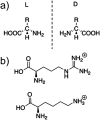
Fig. 1
Fig. 1. (a) The general structure of l- and d-amino acids. (b) The structures of positively charged l-arginine and l-lysine amino acids.
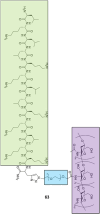
Fig. 2
Fig. 2. Examples of common nanostructures formed via AMP self-assembly: (a) nanofibers (b) nanotubes (c) nanoparticles (d) hydrogels.
Fig. 3
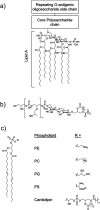
Fig. 3. (a) The structural components of LPS. Molecular composition is known to vary between bacterial species. (b) Lipoteichoic acid structure. (c) An example of a phospholipid structure and the molecular structure of different phospholipid headgroups phosphatidylethanolamine (PE), phosphatidylcholine (PC), phosphatidylglycerol (PG), phosphatidylserine (PS) and cardiolipin.
Fig. 4
Fig. 4. Models by which AMPs elicit antimicrobial activity. (a) If not assembled prior to arriving at the membrane, peptides initially accumulate at the bacterial membrane in a parallel orientation. (b) In the barrel-stave model a pore is formed with peptides in a transmembrane orientation. Hydrophobic amino acids face the lipid membrane and hydrophilic residues line the lumen (the inside of the pore). (c) In the traditional toroidal pore model, peptides bend lipids into the pore; this forms a continuous pore lined by both AMPs and lipid head groups. (d) Disordered toroidal pores are also lined by lipids, however, contain only one or two peptides lining the pore, often located at the edges of the self-assembled structure produced. (e) In the carpet model, AMPs continue to coat the membrane in a parallel formation, forming an extensive layer referred to as a carpet. (f) At high concentrations the carpet can cause disruption via detergent action.
Fig. 5
Fig. 5. The structure of compounds 1–4 as developed by Schnaider et al. and Porter et al.
Fig. 6
Fig. 6. Scanning electron microscopy images of S. aureus. (a–c) Untreated S. aureus (NCTC 10788) biofilm after 24 hours. (d–f) S. aureus (NCTC 10788) biofilm after 24 hour treatment with increasing concentrations of 2. Reproduced with permission from Elsevier.
Fig. 7
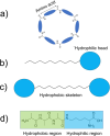
Fig. 7. Example diagrams of common peptidic building blocks. (a) Cyclic peptides formed through amide bond formation of the terminal amino and carboxyl groups of the peptide. (b) Peptide amphiphiles. (c) Bolaamphiphiles displaying the hydrophilic head groups and hydrophobic tail/skeleton. (d) Amphiphilic surfactant-like peptides.
Fig. 8
Fig. 8. Representation of the antiparallel β-sheet formation from cyclic peptides. Alternating d- and l-amino acids displaying a cylindrical structure form antiparallel β-sheets through extensive intermolecular hydrogen bonding. Compounds 5 and 6 synthesised by Fernandez-Lopez et al.
Fig. 9
Fig. 9. The primary structure of compound 7 synthesised by Chang et al. The amphiphilic portion of 7 is shown in the green box, while the heparin-binding Cardin-motif is shown in the blue box.
Fig. 10
Fig. 10. The primary structure of compounds 8 and 9. The alanine residue is displayed in brackets, while the two arginine residues are located on either side of the alanine.
Fig. 11
Fig. 11. Compounds 13–20 developed by Thota et al.
Fig. 12
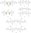
Fig. 12. Compounds 21–26. Green boxes highlight the N–C torsional bond between the phenyl ring and amide carbon. The dotted line indicates hydrogen bonding. R groups in compounds 21–23 were varied in hydrophobicity and charge.
Fig. 13
Fig. 13. Generic structure and R groups for compounds 27–31 developed by Brahmachari et al. Green indicates hydrophobic tail. Blue indicates hydrophilic head.
Fig. 14
Fig. 14. Compounds 32–34 with TBA counter cation. The possible binding modes of the SSA anionic component with (a) phosphatidylcholine (PC), (b) phosphatidylethanolamine (PE) and (c) phosphatidyl glycerol (PG) phospholipid headgroups.
Fig. 15
Fig. 15. Compounds 32–35 developed by Kannan et al.
Fig. 16
Fig. 16. Compounds 39–41 and the resulting Kandinsky circle formed from compound 39 with Cd(ii).
Fig. 17
Fig. 17. Compounds 42–45 synthesised by Sikder et al. The two hydrophilic ends of the molecule (blue), the hydrazide group (red) and the hydrophobic π-conjugated core (green). (a) The resulting polymersome from compounds 42–44. (b) The resulting polymersome from compound 45.
Fig. 18
Fig. 18. The proposed network formation produced through Zn2+ crosslinked compound 47 resulting in hydrogel formation.
Fig. 19
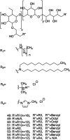
Fig. 19. The structure of compounds 48–55. The first R1 group was varied between C12 and C18. The R3 group on the first quaternary ammonium R2 group was varied between benzyl and octyl groups.
Fig. 20
Fig. 20. Bright field scanning TEM images of the obtained gold NPs synthesised by Piktel et al., which are described by their shape (rod/peanut/star or sphere) and reaction condition (temperature and/or the addition of centrimonium bromide (CTAB). (a) AuR NPs–gold rods, (b) AuP NPs–gold peanuts, (c) AuS NPs–gold stars, (d) AuSph (70C) NPs–gold spherical (70C), (e) AuSph (CTAB) NPs–gold spherical, (f) AuSPph NPs–gold spherical NP. This image has been reproduced with the permission of Nature Scientific Reports.
Fig. 21
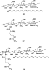
Fig. 21. The three synthetically altered chitosan polymers 56–58 synthesised by Ahmad et al.
Fig. 22
Fig. 22. A cartoon illustrating the triblock copolymer F127 encapsulating (4,7-dibromo-5,6-di(9H-carbazol-9-yl)benzo[c][1,2,5] thiadiazole (DBCz-BT), forming nanoparticles. When irradiated at 410 nm, the encapsulated DBCz-BT can generate active singlet oxygen species.
Fig. 23
Fig. 23. Aspect ratio is an essential feature of antibacterial nanopatterned materials. (a) The difference between high and low aspect ratio nanopatterns. (b) One proposed mechanism of antibacterial action where the 3D patterns rupture the cell wall. (c and d) A second proposed mechanism whereby the bacteria firstly adhere to the nanopattern (c) which results in the tearing of the bacterial cell wall (d).
Fig. 24
Fig. 24. Scanning electron microscopy (1–2) and atomic force microscopy (3–4) images of the (a) micellular (b) cylindrical vertical (c) cylindrical parallel, nanopatterns from Fontelo et al. This image has been reproduced with the permission of Elsevier.
Fig. 25
Fig. 25. The antibacterial efficacy against E. coli DH5α, shown as percentage bacterial efficiency, in response to the different nanopattern pillar lengths, widths and densities from the Michalska et al. study.
Fig. 26
Fig. 26. Stimulus based drug release of an antimicrobial agent from a ‘smart material’. ROS = reactive oxygen species.
Fig. 27
Fig. 27. Articles published on stimuli responsive drug delivery for antibacterial treatment in last 10 years.
Fig. 28
Fig. 28. (a) Thrombin sensitive peptide conjugated silk peptide nanosphere. (b) Activation of nanosphere due to thrombin.
Fig. 29
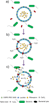
Fig. 29. Activation and action of liposome based Nanoreactor. (a) DSPE-PEG3400, lecithin, lauric acid and stearic acid liposome containing calcium peroxide and rifampicin surrounded by bacteria and alpha toxin; (b) alpha toxin integrates into the membrane, forming a pore through which water enters; (c) water reacts with calcium peroxide to form hydrogen peroxide which decomposes to oxygen gas. The evolution of this gas promotes the release of rifampicin from the liposome.
Fig. 30
Fig. 30. Structure of compound 59.
Fig. 31
Fig. 31. Poly(β-amino ester)-guanidine-phenylboronic acid (PBAE-G-B) cationic polymer based nanoparticle activation. (a) Dextran is cleaved from PBAE-G-B in a low pH environment. (b) Intact nanoparticle in physiological pH environment. (c) Dextran cleavage at low pH causing release of rifampicin and PBAE-G cationic polymer. ROS = reactive oxygen species.
Fig. 32
Fig. 32. Lifecycle of a biofilm (a) free planktonic cells, (b) bacterial cells reversibly adhere to surface, (c) Irreversible attachment occurs with extracellular polymeric substance (EPS) formation, (d) bacterial cells proliferate and biofilm undergoes maturation, (e) mature biofilm is formed with channels for nutrient transportation and removal of metabolic waste. Planktonic cells are also released from biofilm and can go on to restart the cycle.
Fig. 33
Fig. 33. Schematic representation of the composition of biofilm extracellular polymeric substance, including polysaccharides, extracellular DNA, proteins, humic acids and lipids.
Fig. 34
Fig. 34. Changes to poly(di(ethylene glycol)methyl ether methacrylate) (PDEGMA) brush on titanium implant above and below the lower critical solution temperature (LCST). Below the LSCT levofloxacin remains bound to the PDEGMA brushes. Above the LSCT levofloxacin is released from the PDEGMA brushes.
Fig. 35
Fig. 35. Chemical structures of compounds 60–62.
Fig. 36
Fig. 36. Chitosan–polyethylene glycol–LK13 peptide, 63, with chitosan outlined in green, polyethylene glycol in blue, and the LK13 peptide in purple.
Fig. 37
Fig. 37. (top) Curcumin and its attachment point to the poly(lactic-co-glycolic acid)-dextran10) (PLGA-Dex10) copolymer. (bottom) Compound 64 (PLGA-Dex10) copolymer synthesised by Barros et al. Compound 65 is 64 attached to curcumin.
None
Jack A. Doolan
None
George T. Williams
None
Kira L. F. Hilton
None
Rajas Chaudhari
None
John S. Fossey
None
Benjamin T. Goult
None
Jennifer R. Hiscock

References

    1. Prestinaci F. Pezzotti P. Pantosti A. Pathog. Global Health. 2015;109:309–318. doi: 10.1179/2047773215Y.0000000030. - DOI - PMC - PubMed
    1. Charani E. McKee M. Ahmad R. Balasegaram M. Bonaconsa C. Merrett G. B. Busse R. Carter V. Castro-Sanchez E. Franklin B. D. Georgiou P. Hill-Cawthorne K. Hope W. Imanaka Y. Kambugu A. Leather A. J. M. Mbamalu O. McLeod M. Mendelson M. Mpundu M. Rawson T. M. Ricciardi W. Rodriguez-Manzano J. Singh S. Tsioutis C. Uchea C. Zhu N. N. Holmes A. H. Lancet Reg. Health Europe. 2021;7:100161. doi: 10.1016/j.lanepe.2021.100161. - DOI - PMC - PubMed
    1. O'Neill J., Tackling Drug-Resistant Infections Globally: Final Report and Recommendations. Review on Antimicrobial Resistance, Wellcome Trust and HM Government, 2016
    1. Moon S. Williams S. Cullen M. Supportive Cancer Ther. 2006;3:207–216. doi: 10.3816/SCT.2006.n.018. - DOI - PubMed
    1. Nanayakkara A. K. Boucher H. W. Fowler V. G. Jezek A. Outterson K. Greenberg D. E. Ca-Cancer J. Clin. 2021;71:488–504. doi: 10.3322/caac.21697. - DOI - PubMed

MeSH terms

Substances