. 2022 Dec 15;28(24):5405-5418.
doi: 10.1158/1078-0432.CCR-22-0963.

HIF2 Inactivation and Tumor Suppression with a Tumor-Directed RNA-Silencing Drug in Mice and Humans

Affiliations

HIF2 Inactivation and Tumor Suppression with a Tumor-Directed RNA-Silencing Drug in Mice and Humans

Yuanqing Ma et al. Clin Cancer Res. .

Abstract

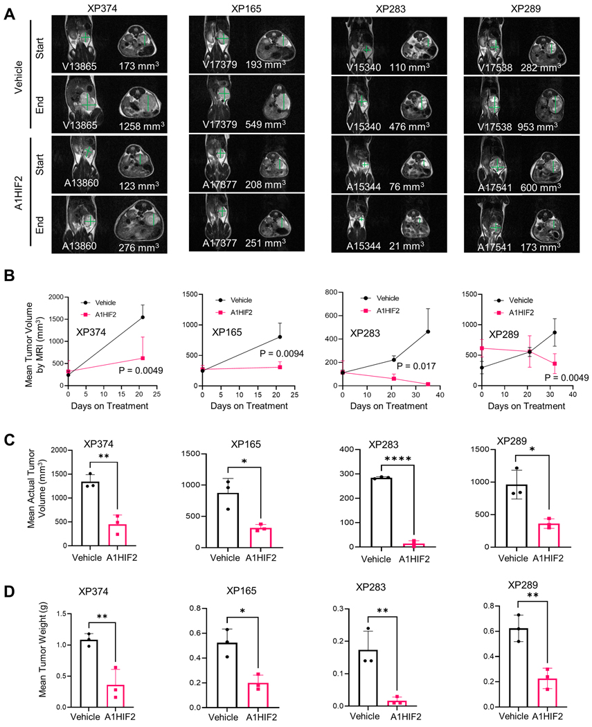
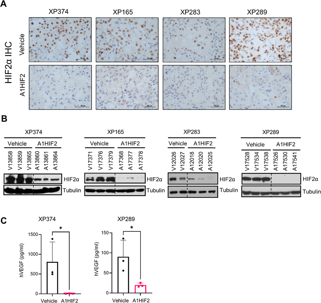
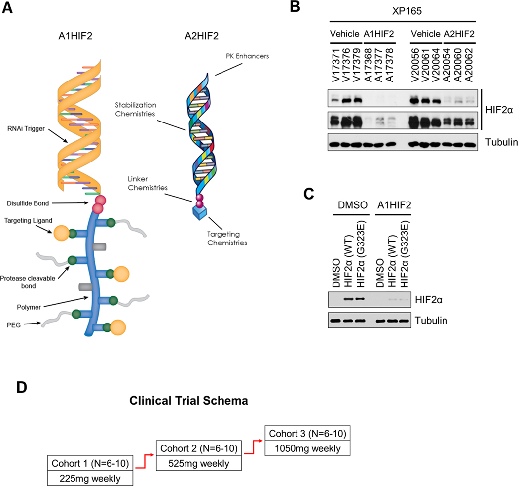
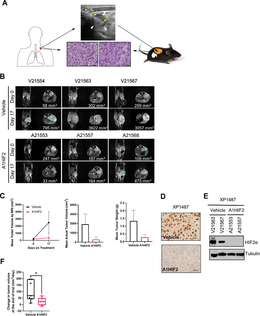
Purpose: HIF2α is a key driver of kidney cancer. Using a belzutifan analogue (PT2399), we previously showed in tumorgrafts (TG) that ∼50% of clear cell renal cell carcinomas (ccRCC) are HIF2α dependent. However, prolonged treatment induced resistance mutations, which we also identified in humans. Here, we evaluated a tumor-directed, systemically delivered, siRNA drug (siHIF2) active against wild-type and resistant-mutant HIF2α.

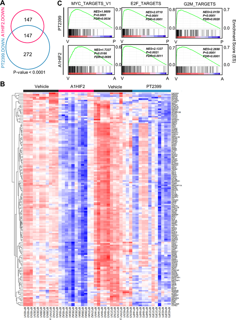
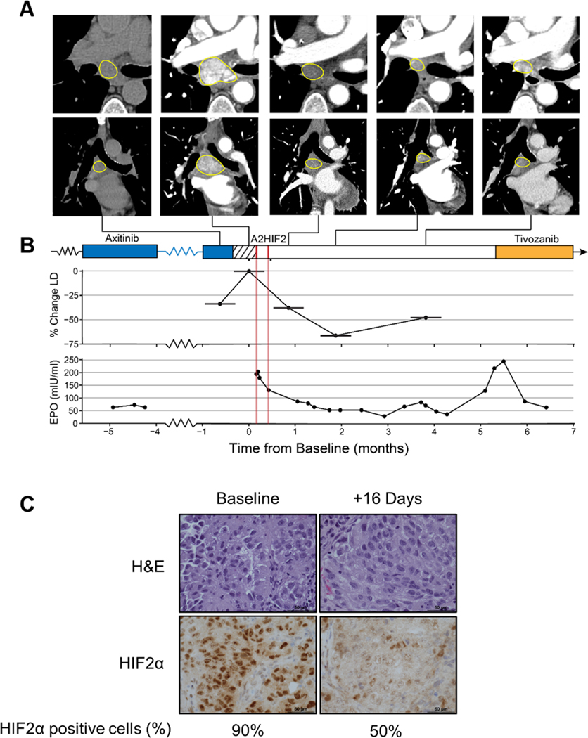
Experimental design: Using our credentialed TG platform, we performed pharmacokinetic and pharmacodynamic analyses evaluating uptake, HIF2α silencing, target gene inactivation, and antitumor activity. Orthogonal RNA-sequencing studies of siHIF2 and PT2399 were pursued to define the HIF2 transcriptome. Analyses were extended to a TG line generated from a study biopsy of a siHIF2 phase I clinical trial (NCT04169711) participant and the corresponding patient, an extensively pretreated individual with rapidly progressive ccRCC and paraneoplastic polycythemia likely evidencing a HIF2 dependency.

Results: siHIF2 was taken up by ccRCC TGs, effectively depleted HIF2α, deactivated orthogonally defined effector pathways (including Myc and novel E2F pathways), downregulated cell cycle genes, and inhibited tumor growth. Effects on the study subject TG mimicked those in the patient, where HIF2α was silenced in tumor biopsies, circulating erythropoietin was downregulated, polycythemia was suppressed, and a partial response was induced.

Conclusions: To our knowledge, this is the first example of functional inactivation of an oncoprotein and tumor suppression with a systemic, tumor-directed, RNA-silencing drug. These studies provide a proof-of-principle of HIF2α inhibition by RNA-targeting drugs in ccRCC and establish a paradigm for tumor-directed RNA-based therapeutics in cancer.


Figures

Fig. 1.
Fig. 1.. Tumor growth inhibition by siHIF2 in ccRCC TGs.
A. Representative T2-weighted MRI images at baseline and after administration of A1HIF2 with corresponding tumor volume quantitation. B. Tumor volume by MRI (n=3 per arm) at baseline and at the end of treatment. C. Actual tumor volume measurements at the end of treatment. D. Tumor weight at the end of treatment. *, p < 0.05; **, p<0.01; ****, p < 0.0001. Error bars represent standard deviation.
Fig. 2.
Fig. 2.. HIF2α silencing and VEGF downregulation by siHIF2.
A. Representative immunohistochemistry images illustrating HIF2α protein depletion by A1HIF2 in TGs. B. Western blot analyses of HIF2α in A1HIF2 treated TG-bearing mice. C. Human VEGF ELISA of circulating tumor-produced VEGF in A1HIF2 or vehicle treated mice. *, p < 0.05. Error bars represent standard deviation.
Fig. 3.
Fig. 3.. Orthogonal analyses of siHIF2 and PT2399 in TGs define the HIF2 transcriptome in ccRCC.
A. Venn diagram showing shared downregulated genes (FDR < 0.05 and LogFC < −1) by A1HIF2 and PT2399 in TGs (11 vehicle- and 11 A1HIF2-treated tumors from XP165, XP283, XP289, and XP374; and 12 vehicle- and 12 PT2399-treated tumors from XP144, XP164, XP373, XP374, and XP453). B. Heatmap of the overlapping 147 significantly downregulated genes. C. Unsupervised GSEA of overlapping 147 genes showing convergence on MYC, E2F, and G2M targets.
Fig. 4.
Fig. 4.. First (A1HIF2) and second generation siHIF2 (A2HIF2).
A. Schematic illustration of A1HIF2 and A2HIF2, which share the same RNAi trigger. B. Comparative analyses of HIF2α depletion by A1HIF2 and A2HIF2. C. Depletion of ectopically expressed HIF2α with acquired resistance mutation (G323E) in HEK293T by siHIF2 (A1HIF2). D. Clinical trial schema of A2HIF2 (NCT04169711).
Fig. 5.
Fig. 5.. HIF2α inhibition and tumor suppression by siHIF2 in TG from clinical trial participant (106–00C).
A. Schematic illustrating orthotopic TG line generation from ultrasound-guided biopsy (yellow arrows) of a supraclavicular lymph node (white arrowheads) from 106–00C (day 16) along with representative H&E images of the patient core biopsy and corresponding TG (XP1487). B. MRI images of orthotopic tumor-bearing TGs treated with A1HIF2 (or vehicle). C. Tumor volume and tumor weight analyses. D. Representative HIF2α immunohistochemistry images from A1HIF2-treated or control TGs. E. Western blot analysis of HIF2α protein in TGs treated with A1HIF2 or vehicle. F. Integrated analyses of two A1HIF2 trials in TGs from 106–00C (see also Sup Fig. 8). *, p < 0.05. Error bars represent standard deviation.
Fig. 6.
Fig. 6.. HIF2α downregulation, Epo suppression and tumor growth inhibition by siHIF2 in clinical trial participant (106–00C).
A. Axial (top) and coronal (bottom) CT images of subcarinal lymph node target lesion showing rapid progression during 2-week washout period and deep prolonged response after two doses of A2HIF2. While the first CT scan was performed without iodinated contrast, marked decrease in lymph node enhancement between baseline (second) CT and subsequent CTs illustrates profound antiangiogenic effect. B. Treatment timeline, tumor change (LD, longest diameter) and erythropoietin (EPO) levels prior to and following A2HIF2 administration (red lines). C. H&E and HIF2α immunohistochemistry of lymph node biopsy at baseline and 16 days after treatment onset with percent of tumor cells positively staining for HIF2α.

References

    1. Shen C. and Kaelin WG Jr., The VHL/HIF axis in clear cell renal carcinoma. Semin Cancer Biol, 2013. 23(1): p. 18–25. - PMC - PubMed
    1. Latif F, et al., Identification of the von Hippel-Lindau disease tumor suppressor gene. Science, 1993. 260(5112): p. 1317–20. - PubMed
    1. Gnarra JR, et al., Mutations of the VHL tumour suppressor gene in renal carcinoma. Nat Genet, 1994. 7(1): p. 85–90. - PubMed
    1. Herman JG, et al., Silencing of the VHL tumor-suppressor gene by DNA methylation in renal carcinoma. Proc Natl Acad Sci U S A, 1994. 91(21): p. 9700–4. - PMC - PubMed
    1. Nickerson ML, et al., Improved identification of von Hippel-Lindau gene alterations in clear cell renal tumors. Clin Cancer Res, 2008. 14(15): p. 4726–34. - PMC - PubMed

Publication types

MeSH terms