. 2022 Oct 3;18(10):e1010855.
doi: 10.1371/journal.ppat.1010855. eCollection 2022 Oct.

Human neutrophil IL1β directs intestinal epithelial cell extrusion during Salmonella infection

Affiliations

Human neutrophil IL1β directs intestinal epithelial cell extrusion during Salmonella infection

Anna-Lisa E Lawrence et al. PLoS Pathog. .

Abstract

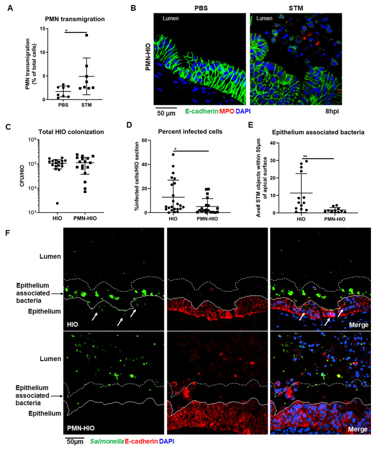
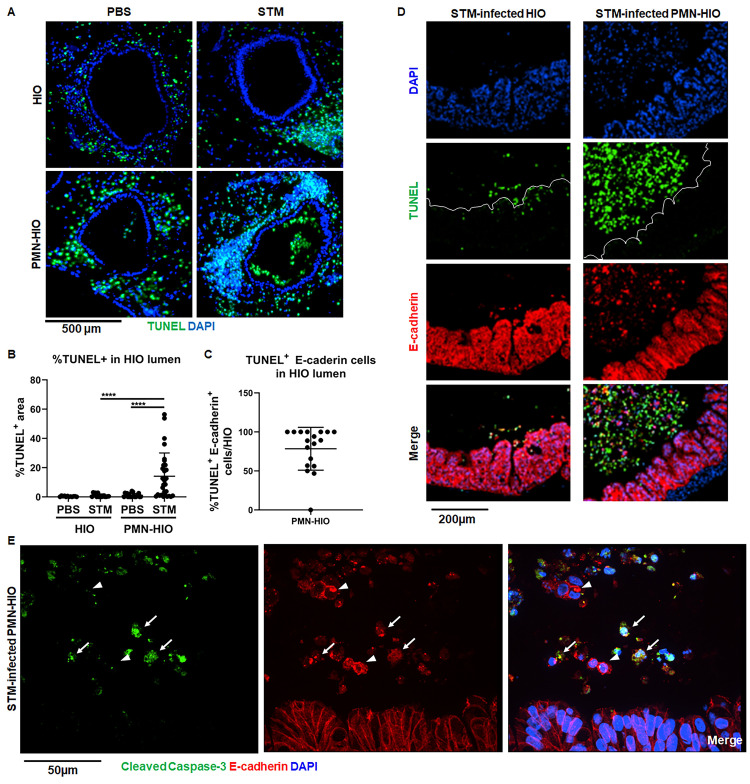
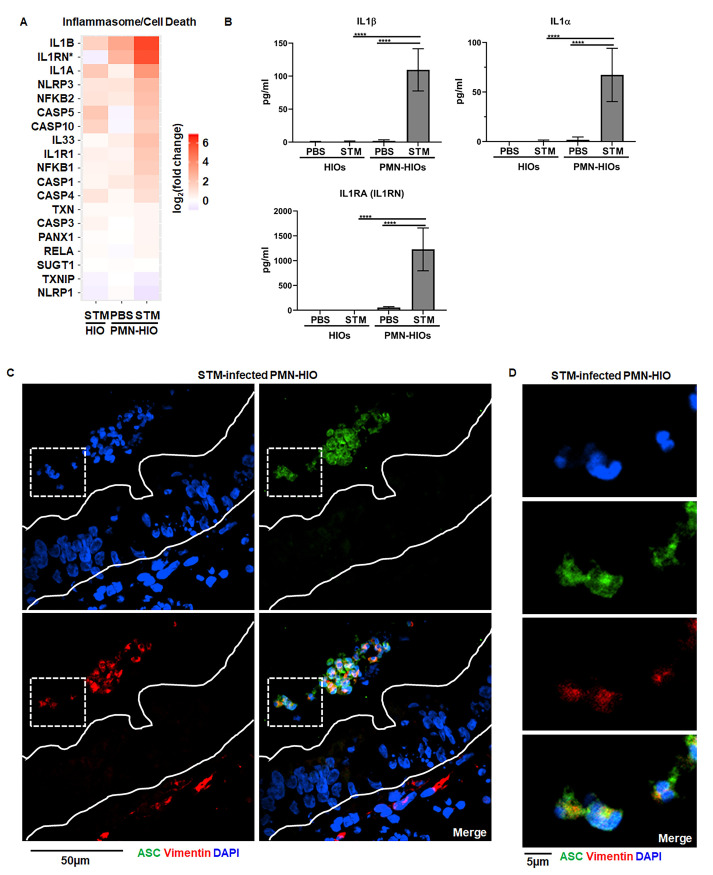
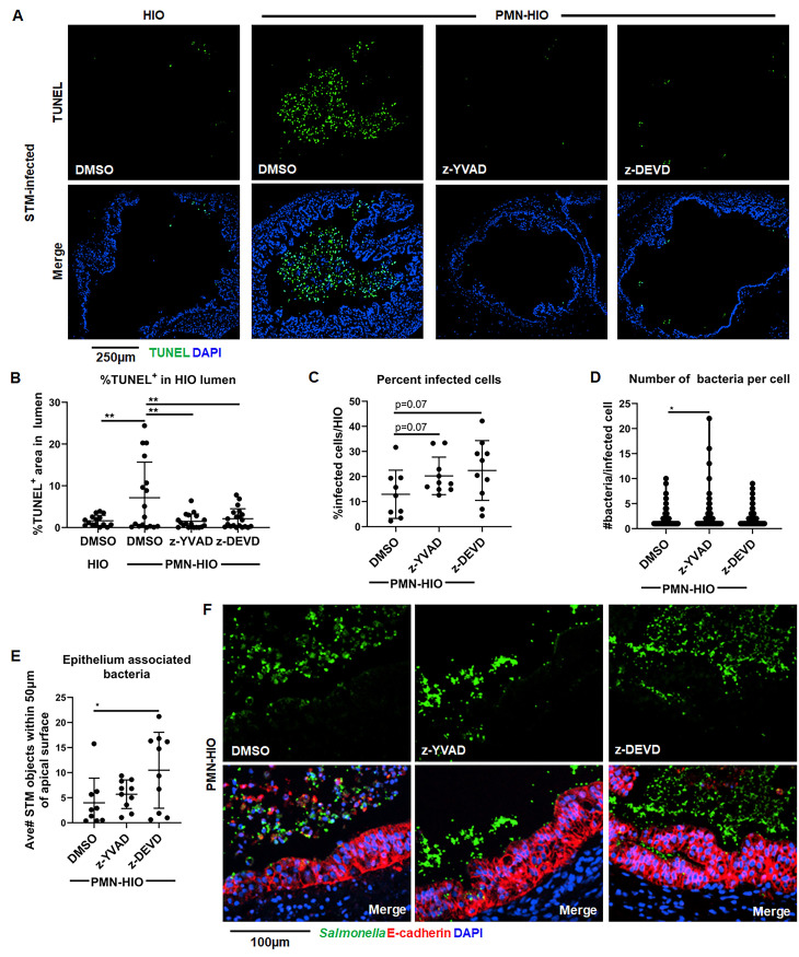
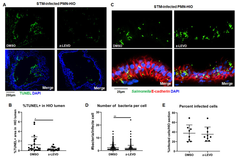
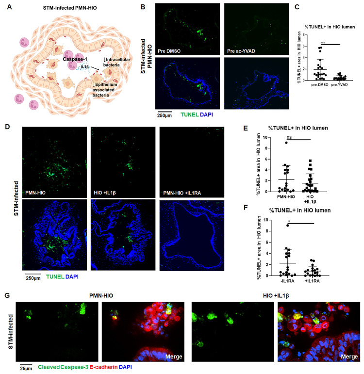
Infection of the human gut by Salmonella enterica Typhimurium (STM) results in a localized inflammatory disease that is not mimicked in murine infections. To determine mechanisms by which neutrophils, as early responders to bacterial challenge, direct inflammatory programming of human intestinal epithelium, we established a multi-component human intestinal organoid (HIO) model of STM infection. HIOs were micro-injected with STM and seeded with primary human polymorphonuclear leukocytes (PMN-HIOs). PMNs did not significantly alter luminal colonization of Salmonella, but their presence reduced intraepithelial bacterial burden. Adding PMNs to infected HIOs resulted in substantial accumulation of shed TUNEL+ epithelial cells that was driven by PMN Caspase-1 activity. Inhibition of Caspases-1, -3 or -4 abrogated epithelial cell death and extrusion in the infected PMN-HIOs but only Caspase-1 inhibition significantly increased bacterial burden in the PMN-HIO epithelium. Thus, PMNs promote cell death in human intestinal epithelial cells through multiple caspases as a protective response to infection. IL-1β was necessary and sufficient to induce cell shedding in the infected HIOs. These data support a critical innate immune function for human neutrophils in amplifying cell death and extrusion of human epithelial cells from the Salmonella-infected intestinal monolayer.


Conflict of interest statement

The authors have declared that no competing interests exist.

Figures

Fig 1
Fig 1. PMNs migrate into the lumen during infection and reduce both the number of infected epithelial cells and the association of bacteria with the epithelial surface.
A. Transmigration of PMNs into the lumen of HIOs was quantified using flow cytometry. HIOs were microinjected with STM or PBS and co-cultured with CFSE-labeled PMNs for 8h. PMNs-HIOs were washed to remove any unassociated PMNs, dissociated into a single cell suspension and subjected to flow cytometry. Percentage of PMNs relative to total cells acquired per PMN-HIO was determined by FlowJo software. B. Immunofluorescent staining of HIOs microinjected with PBS or STM and co-cultured with PMNs. E-cadherin (green) marks the epithelial lining, MPO (red) is specific to PMNs and DNA is stained with DAPI. C. Total bacterial burden per HIO or PMN-HIO was enumerated at 8hpi. Individual HIOs were removed from Matrigel, washed with PBS and homogenized in PBS. Serial dilutions were plated on LB agar to enumerate bacterial burden. D. Quantitation of percent infected cells/HIO or PMN-HIO based on 3 fields per view per HIO. E. Quantitation of epithelium associated bacteria. Number of bacteria within 50μm of the apical epithelial surface based on E-cadherin staining were counted and normalized per 100μm distance. F. Representative immunofluorescent staining of HIOs and PMN-HIOs infected with STM. Salmonella is stained in green, E-cadherin to mark epithelial cells is shown in red, and DNA stained with DAPI in blue. White arrows point to infected cells. Graphs show the mean and SD of n≥ 10 HIOs represented by dots from at least two independent experiments. Outliers were removed using the ROUT method with Q = 0.1%. Unless otherwise stated, significance was determined by Mann-Whitney test with *p<0.05, **p<0.01.
Fig 2
Fig 2. PMNs induce epithelial cell death and shedding during Salmonella infection.
A. Immunofluorescent images of TUNEL staining of histology sections of HIOs and PMN-HIOs injected with PBS or STM at 8hpi. B. Quantitation of TUNEL positive cells in the lumen of HIOs and PMN-HIOs from (A). Graphs show the mean and SD of HIOs from 2 independent experiments with n>12 HIOs per group. C. Quantitation of percent of TUNEL-positive epithelial cells in HIO lumen. The percentage of TUNEL-positive cells that stained positive for E-cadherin in the HIO lumen were assessed. D. Representative confocal microscopy images of histology sections from STM-injected HIOs or PMN-HIOs at 8h. Sections were co-stained with TUNEL (green), epithelial cell marker E-cadherin (red), and DNA marker DAPI (blue). E. Confocal microscopy images of histology sections of HIOs and PMN-HIOs that were stained for E-cadherin (red), cleaved Caspase-3 (green), and DNA (blue). Arrows point to cleaved Caspase-3 positive epithelial cells whereas arrowheads point to cleaved Caspase-3 negative luminal epithelial cells. Outliers were removed using the ROUT method with Q = 0.1%. Significance was determined via one-way ANOVA with post-Tukey’s test for multiple comparisons where ****p<0.0001.
Fig 3
Fig 3. Inflammasome activation and IL-1 production is mediated by PMNs during infection.
A. Gene expression data presented as log2(fold change) relative to PBS-injected HIOs for genes involved in inflammasome/cell death signaling. All genes are significantly changed from PBS-injected HIOs in at least one condition with p-adjusted value <0.05. B. Cytokine levels in culture media of HIOs and PMN-HIOs were quantified using ELISA. Graphs indicate the mean of n = 4 biological replicates +/- SD from media sampled at 8hpi with 5 HIOs or PMN-HIOs per well. C. Immunofluorescent staining of histology sections of PMN-HIOs. Sections were stained for ASC expression (green), Vimentin (red) to mark PMNs and mesenchymal cells, and DNA (blue) was labeled with DAPI. D. Zoom of (C) showing luminal ASC-positive cells (green) with multilobed PMN nuclei. Statistical significance was determined by 2-way ANOVA where *p<0.05, ***p<0.001, ****p<0.0001.
Fig 4
Fig 4. Caspase-1 and Caspase-3 inhibition reduces shedding of infected epithelial cells in the lumen of PMN-HIOs and differentially affect bacterial burden and bacterial association with the epithelium.
A. Representative fluorescence microscopy images of TUNEL staining of HIO and PMN-HIO histology sections. HIOs were microinjected with STM and either cultured alone or co-cultured with PMNs in the presence of inhibitors for Caspase-1 (z-YVAD), Caspase-3 (z-DEVD), or DMSO control. B. Quantitation of the percent of lumen filled with TUNEL-positive cells of STM-infected HIOs or PMN-HIOs with indicated treatments. C. Quantitation of the percent of infected cells per HIO based on 3 fields per view per HIO. D. Quantitation of number of bacteria per infected cell in PMN-HIOs based on 3 fields per view per HIO. E. Quantitation of epithelium associated bacteria. Number of bacteria within 50μm of the apical epithelial surface were counted and normalized per 100μm distance. F. Fluorescent microscopy images of STM-infected PMN-HIO histology sections. Samples were stained for Salmonella (green), E-cadherin (red), and DAPI (blue). Unless otherwise stated, graphs show the mean +/-SD of n≥ 10 HIOs represented by dots from at least two independent experiments. Outliers were removed using the ROUT method with Q = 0.1%. Significance was determined by one-way ANOVA with post-Tukey’s test for multiple comparisons where *p<0.05, **p<0.01.
Fig 5
Fig 5. Caspase-4 regulates cell death in STM-infected PMN-HIOs.
A. Representative fluorescence microscopy images of TUNEL staining of PMN-HIO frozen histology sections. HIOs were microinjected with STM and co-cultured with PMNs in the presence of inhibitors for Caspase-4 (z-LEVD) (20 μM), or DMSO control. B. Quantitation of the percent of lumen filled with TUNEL-positive cells of STM-infected PMN-HIOs with indicated treatments. C. Fluorescent microscopy images of STM-infected PMN-HIO frozen histology sections. Samples were stained for Salmonella (green), E-cadherin (red), and DAPI (blue). D. Quantitation of the number of bacteria per infected cell. E. Quantitation of the percent of infected cells per HIO based on 3 fields per view per HIO. Unless otherwise stated, graphs show the mean +/-SD of n≥ 10 HIOs represented by dots from at least two independent experiments. Outliers were removed using the ROUT method with Q = 0.1%. Significance was determined by unpaired t-test where *p<0.05, **p<0.01.
Fig 6
Fig 6. IL-1 signaling mediates shedding of epithelial cells in STM-infected PMN-HIOs.
A. Cartoon model showing Caspase-1 activation in PMNs leading to IL-1β production which regulates cell death in STM-infected PMN-HIOs. B. Representative fluorescence microscopy images of TUNEL staining of PMN-HIO frozen histology sections. PMNs were pre-treated with Caspase-1 inhibitor (ac-YVAD) for 1h prior to co-culturing with STM-infected HIOs. C. Quantitation of the percent of lumen filled with TUNEL-positive cells of STM-infected PMN-HIOs with PMNs pretreated with either DMSO or ac-YVAD. D. Representative fluorescence microscopy images of TUNEL staining of HIO or PMN-HIO frozen histology sections. HIOs were microinjected with STM or STM +IL1β for 2h and then where indicated HIOs were co-cultured with PMNs alone or cultured with PMNs and treated with IL1RA (1 μg/ml). E. Quantitation of the percent of lumen filled with TUNEL-positive cells of STM-infected HIOs either co-cultured with PMNs or co-microinjected with recombinant IL1β. F. Quantitation of the percent of lumen filled with TUNEL-positive cells of STM-infected PMN-HIOs treated with or without IL1RA. G. Confocal microscopy images of frozen histology sections of STM-infected HIOs microinjected with recombinant IL1β or STM-infected PMN-HIOs that were stained for E-cadherin (red), cleaved Caspase-3 (green), and DNA (blue). Unless otherwise stated, graphs show the mean +/-SD of n≥ 10 HIOs represented by dots from at least two independent experiments. Outliers were removed using the ROUT method with Q = 0.1%. Significance was determined by unpaired t-test where *p<0.05, ***p<0.001.

References

    1. Salmonella Homepage. 27 Jul 2022 [cited 27 Jul 2022]. Available: https://www.cdc.gov/salmonella/index.html
    1. Baird-Parker AC. Foodborne salmonellosis. Lancet. 1990;336: 1231–1235. doi: 10.1016/0140-6736(90)92844-8 - DOI - PubMed
    1. Crowley SM, Vallance BA, Knodler LA. Noncanonical inflammasomes: Antimicrobial defense that does not play by the rules. Cell Microbiol. 2017;19. doi: 10.1111/cmi.12730 - DOI - PubMed
    1. Sellin ME, Müller AA, Felmy B, Dolowschiak T, Diard M, Tardivel A, et al.. Epithelium-intrinsic NAIP/NLRC4 inflammasome drives infected enterocyte expulsion to restrict Salmonella replication in the intestinal mucosa. Cell Host Microbe. 2014;16: 237–248. doi: 10.1016/j.chom.2014.07.001 - DOI - PubMed
    1. Rauch I, Deets KA, Ji DX, von Moltke J, Tenthorey JL, Lee AY, et al.. NAIP-NLRC4 inflammasomes coordinate intestinal epithelial cell expulsion with eicosanoid and IL-18 release via activation of Caspase-1 and -8. Immunity. 2017;46: 649–659. doi: 10.1016/j.immuni.2017.03.016 - DOI - PMC - PubMed

Publication types