

Review
doi: 10.1101/cshperspect.a041087.
The Burial: Clearance and Consequences
Affiliations
- PMID: 36192119
- PMCID: PMC9524284
- DOI: 10.1101/cshperspect.a041087
Item in Clipboard
Review
The Burial: Clearance and Consequences
Cold Spring Harb Perspect Biol.
.
No abstract available
Figures
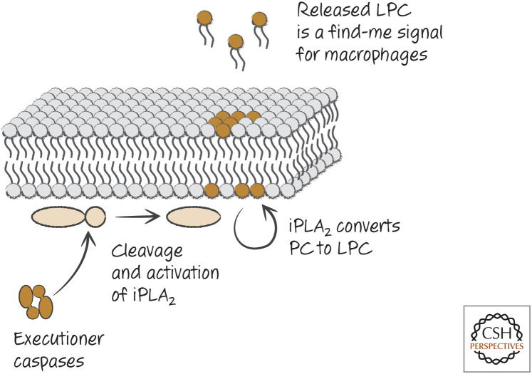
Executioner caspases induce production of the “find-me” signal LPC. LPC, lysophosphatidylcholine; iPLA2, calcium-independent phospholipase A2; PC, phosphatidylcholine.
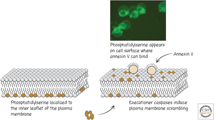
Plasma membrane scrambling and its detection in dying cells. Phosphatidylserine “scrambled” from the inner to the outer membrane can be detected by probes, such as fluorescently labeled annexin V. (Reprinted from Brumatti et al. 2008, ©2008 with permission from Elsevier.)
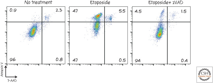
Detection of phosphatidylserine exposure by fluorescence-activated cell sorting (FACS). Cells were treated with the chemotherapy agent etoposide to induce apoptosis (± the caspase inhibitor zVAD-fmk) and then stained with annexin V (coupled with a fluorescent dye) and the vital dye 7-AAD. The fluorescence intensity of each cell is represented by a dot. Notice that the cell population becomes positive for annexin V before membrane integrity (measured by 7-AAD) is lost. This annexin V staining, and by implication phosphatidylserine exposure, is dependent on caspases, as treatment with the inhibitor zVAD largely prevented it. 7-AAD, 7-amino actinomycin D; zVAD, zVAD-fmk.
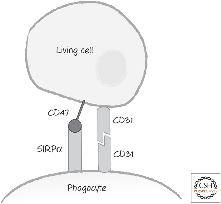
“Don't-eat-me” signals. These include the leukocyte surface antigen CD47, recognized by phagocyte cell-surface receptor SIRPα, and the homotypic interaction between CD31 molecules. SIRPα, signal-regulatory protein alpha.
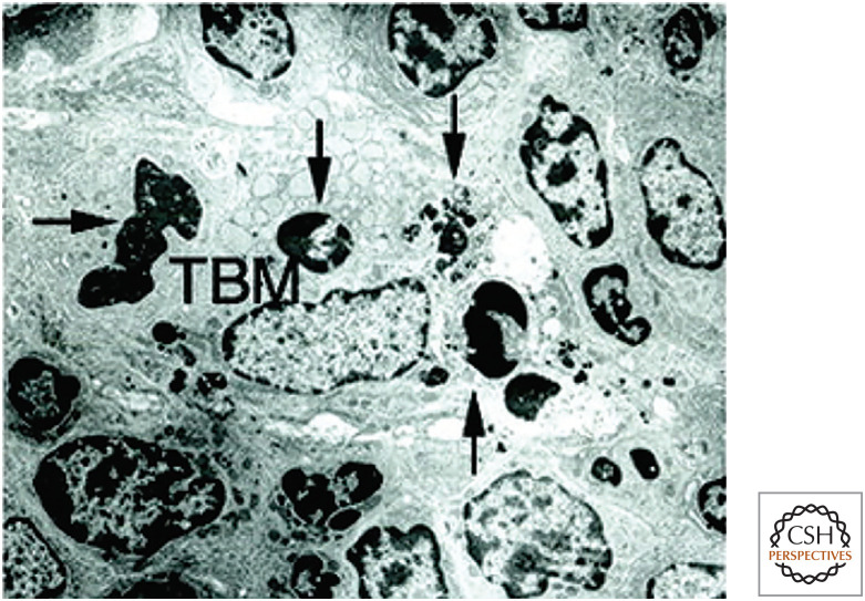
A single tingible body macrophage (TBM) in a germinal center engulfing four apoptotic cells (arrows). (Reprinted from Hanayama et al. 2004, ©2004 with permission from AAAS.)
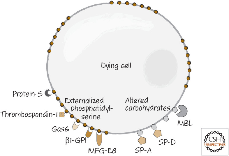
Bridge molecules are soluble molecules that bind to dying cells because of the changes in the plasma membrane. MBL, mannose-binding lectin; MFG-E8, lactadherin.
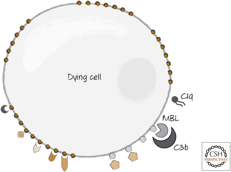
Complement components C1q and C3b can also bind to dying cells to act as bridge molecules.
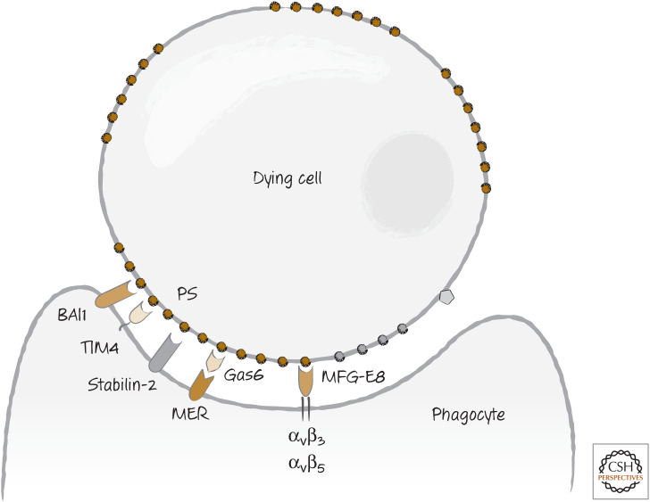
Recognition of phosphatidylserine (PS), exposed on dying cells, by phagocytes occurs either directly (e.g., for BAI1, stabilin 2, TIM4) or indirectly through bridge molecules (e.g., for MER, integrins).
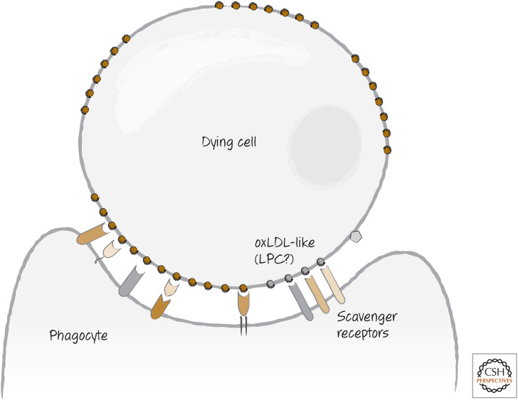
Scavenger receptors recognize dying cells expressing, for example, altered carbohydrates and exposed phosphatidylserine.
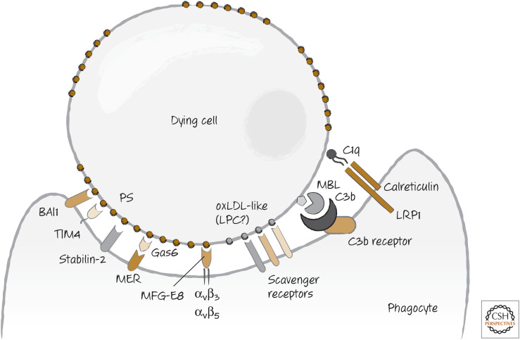
The array of “bind-me” receptors that can directly, or indirectly through bridge molecules, bind to dying cells.
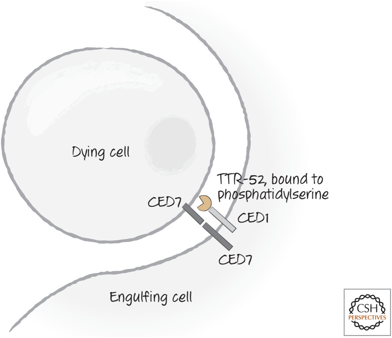
Recognition receptors CED1 and CED7 in nematodes. CED7 is important in both the dying and the engulfing cells, whereas CED1 is important in the engulfing cell. CED1 can bind to a bridging molecule, TTR-52, which recognizes phosphatidylserine and is required for clearance of dying cells. There is at least one additional receptor (not illustrated) involved in recognition and engulfment of dying cells.
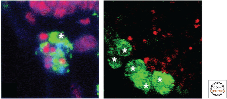
The Drosophila Croquemort mutant has defects in engulfment of dying cells. Here, phagocytic cells (green, with white stars) take up small dying cells (red) in wild-type flies (left) but not in mutants unable to express Croquemort (right). (Franc et al. 1999.)
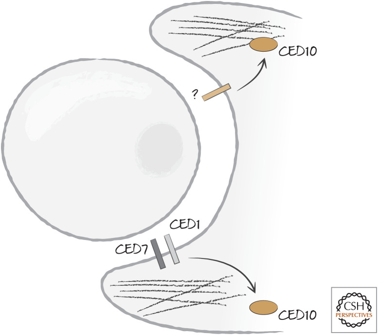
Two pathways converge on the Ras-related protein CED10 (Rac1) to induce actin reorganization and phagocytosis. Although one pathway is linked to CED1 and CED7, the other pathway is initiated in the nematode Caenorhabditis elegans by an unknown receptor.
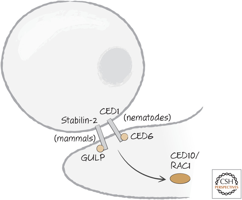
CED1 (nematodes) and stabilin-2 (mammals) engage an adapter molecule, CED6 or GULP, respectively, following recognition of dying cells. CED6/GULP activates CED10/RAC1 by an unknown mechanism.
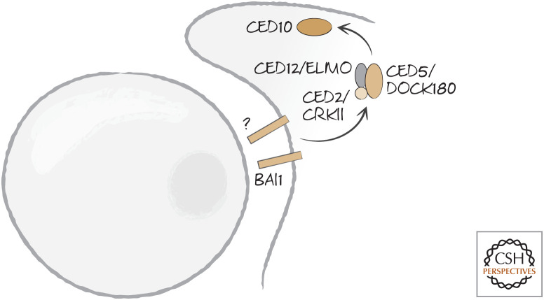
BAI1 in mammals, and an unknown receptor in nematodes, engage a signaling complex (comprising CRKII–DOCK180–ELMO–Rac1 or CED2–CED5–CED12–CED10, respectively) following recognition of dying cells.
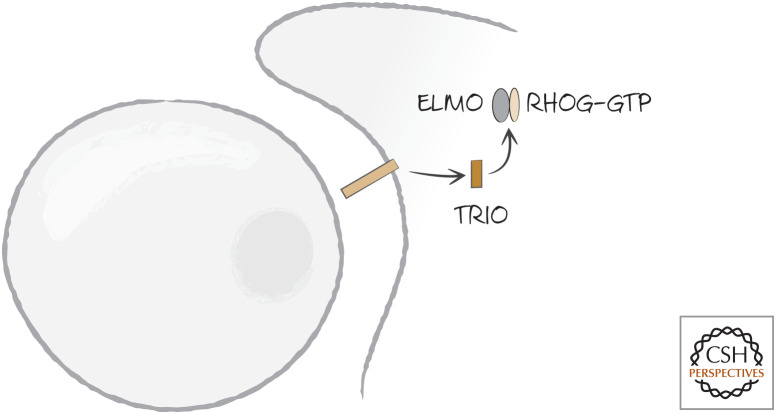
The phosphatidylserine-binding receptor BAI1 (and other receptors) in mammals induce the GDP–GTP exchange factor TRIO, which activates the small GTPase RHOG, converting it to the GTP-bound active form.
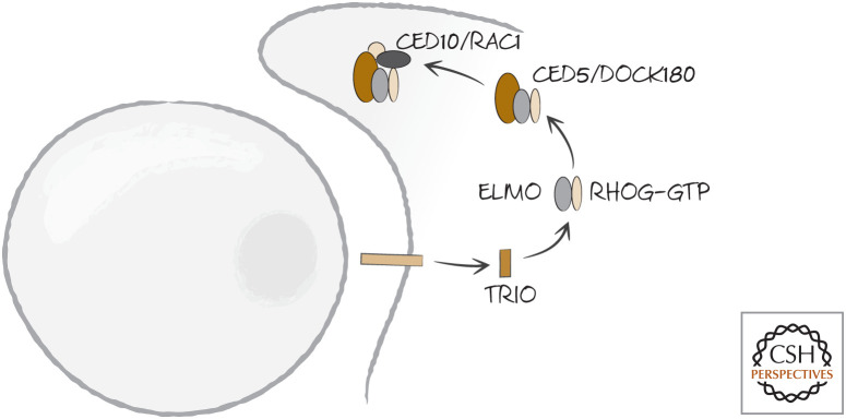
ELMO recruits DOCK180, which binds to RAC1, leading to the activation of this small GTPase. Engulfment by the phagocyte then proceeds.
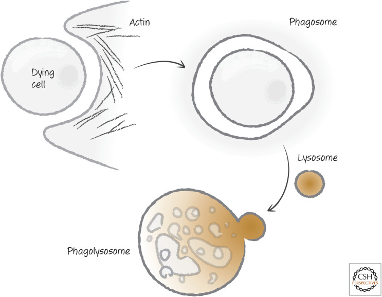
The phagosome and degradation. The phagocyte envelops the dying cell, forming an internal phagosome that, on rearrangement of the actin cytoskeleton and fusion with a lysosome, forms a phagolysosome, where disposal of the dying cell takes place.
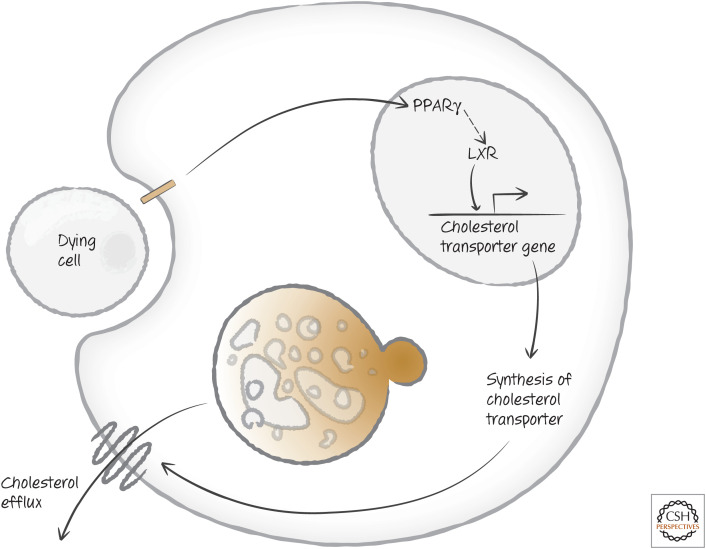
Uptake of dying cells induces cholesterol management. Activation of the peroxisome proliferator-activated receptor γ (PPARγ) induces the expression of the nuclear oxysterols receptor LXR-alpha (NR1H3), which in turn induces the expression of a cholesterol transporter operating at the plasma membrane.
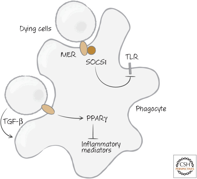
Apoptotic cells can inhibit inflammation. Mer, tyrosine-protein kinase Mer; PPAR γ, peroxisome proliferator-activated receptor γ; SOCS1, suppressor of cytokine signaling 1; TGF-β, transforming growth factor beta; TLR, Toll-like receptor.
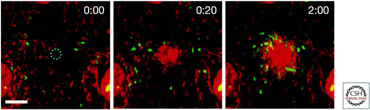
Neutrophils swarm to sites of cell damage. A single cell was damaged by laser light (dashed circle). Within seconds, the neutrophils (red) swarm to the site, followed by macrophages (green). The elapsed time is shown (minutes:seconds).
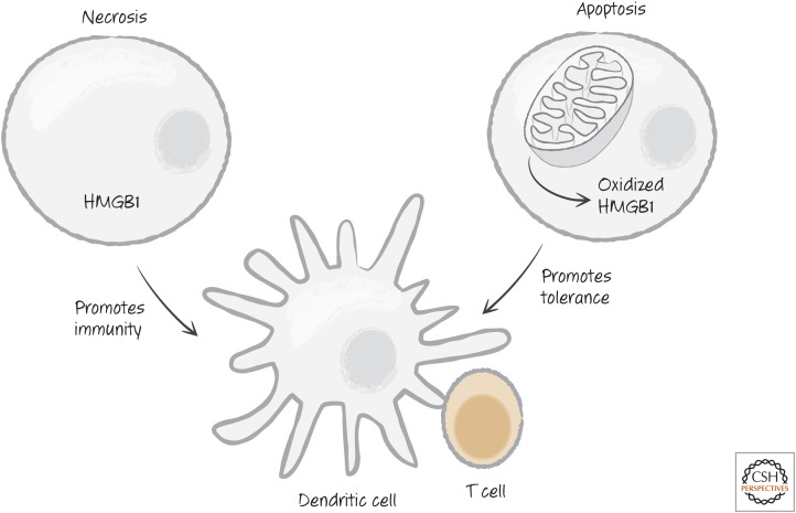
The high-mobility group protein B1 (HMGB1) in immunity versus tolerance. Apoptotic cells, unlike necrotic cells, end up oxidizing HMGB1, preventing it from promoting an immune response.
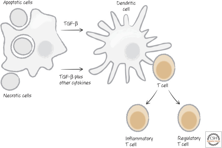
Dying cells influence T-cell functional differentiation. TGF-β, transforming growth factor beta.
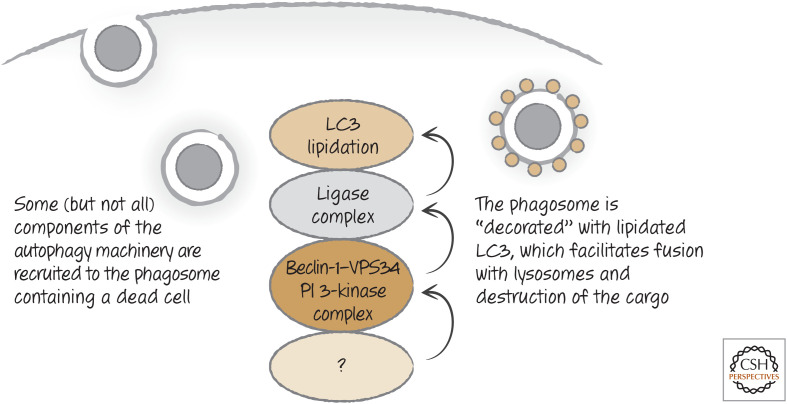
LC3-associated phagocytosis occurs on engulfment of dying cells. As the phagosome forms, an unknown signal is generated that recruits a complex containing beclin-1 and the phosphoinositide 3-kinase VPS34, and other molecules distinct from those involved in autophagy. As a result, the ligase complex is recruited and LC3 is conjugated to lipids in the phagosome membrane. This facilitates fusion with lysosomes and the degradation of the corpse.
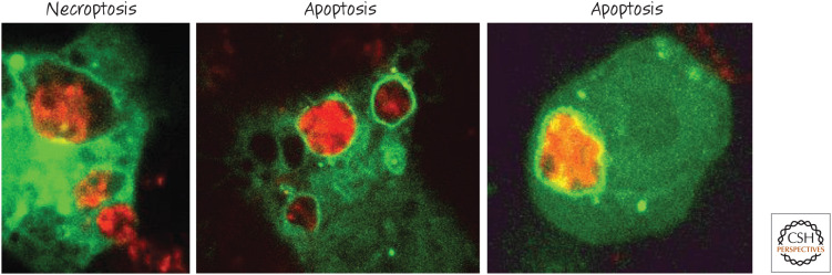
LC3-associated phagocytosis occurs on engulfment of dying cells. Macrophages expressing an LC3 fused to green-fluorescent protein (GFP) were cocultured with cells (stained red) that were dying by means of different mechanisms. Note the ring of green LC3 around the corpse-containing phagosomes. (Provided by Jennifer Martinez and Clifford Guy, Department of Immunology, St. Jude Children's Research Hospital.)
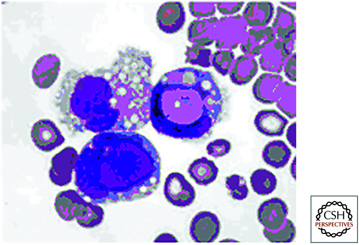
LE bodies. These are apoptotic cells in the circulation and are diagnostic of systemic lupus erythematosus (SLE). (Reprinted from Holman 1951, with permission from BMJ Publishing Group Ltd.)
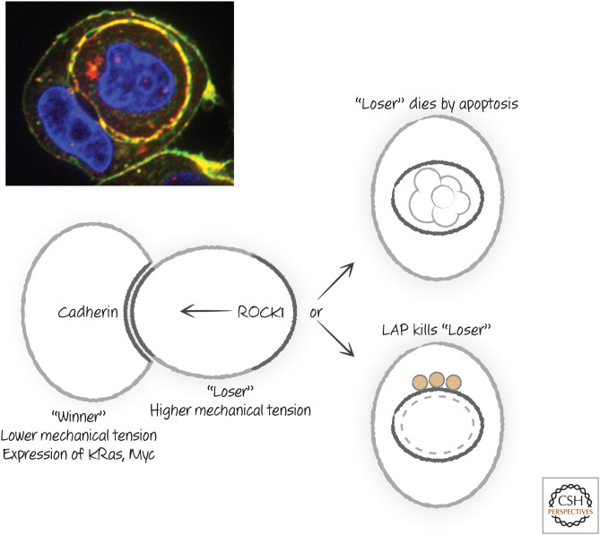
Pathways and molecules of cell death by entosis. LAP, LC3-associated phagocytosis. (Michael Overholter, unpublished.)
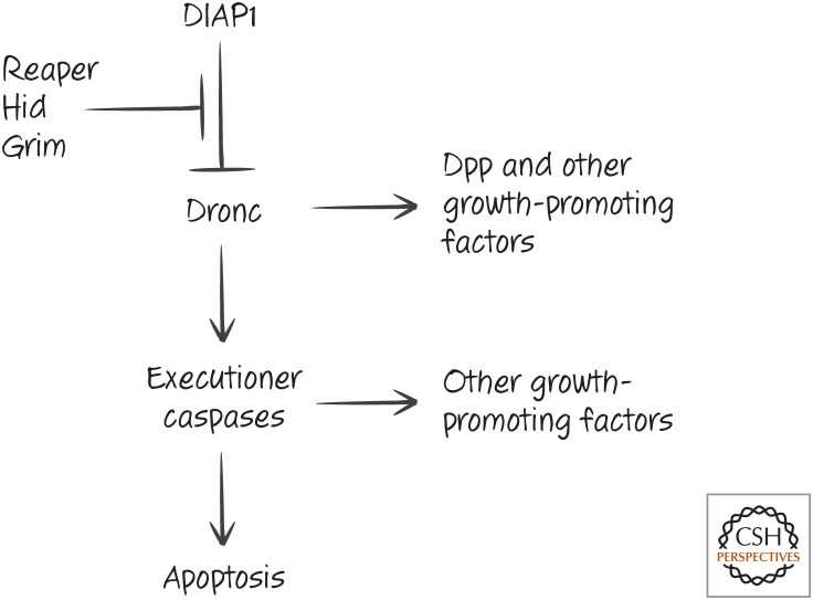
Caspase activation promotes compensatory proliferation in the fruit fly Drosophila. DIAP1, death-associated inhibitor of apoptosis 1; Dpp, Decapentaplegic.
References
Publication types
MeSH terms
LinkOut - more resources
Full Text Sources