Review
. 2022 Dec;26(12):1133-1152.
doi: 10.1016/j.tics.2022.09.006. Epub 2022 Sep 30.

A role for the claustrum in cognitive control

Affiliations
Review

A role for the claustrum in cognitive control

Maxwell B Madden et al. Trends Cogn Sci. 2022 Dec.

Abstract

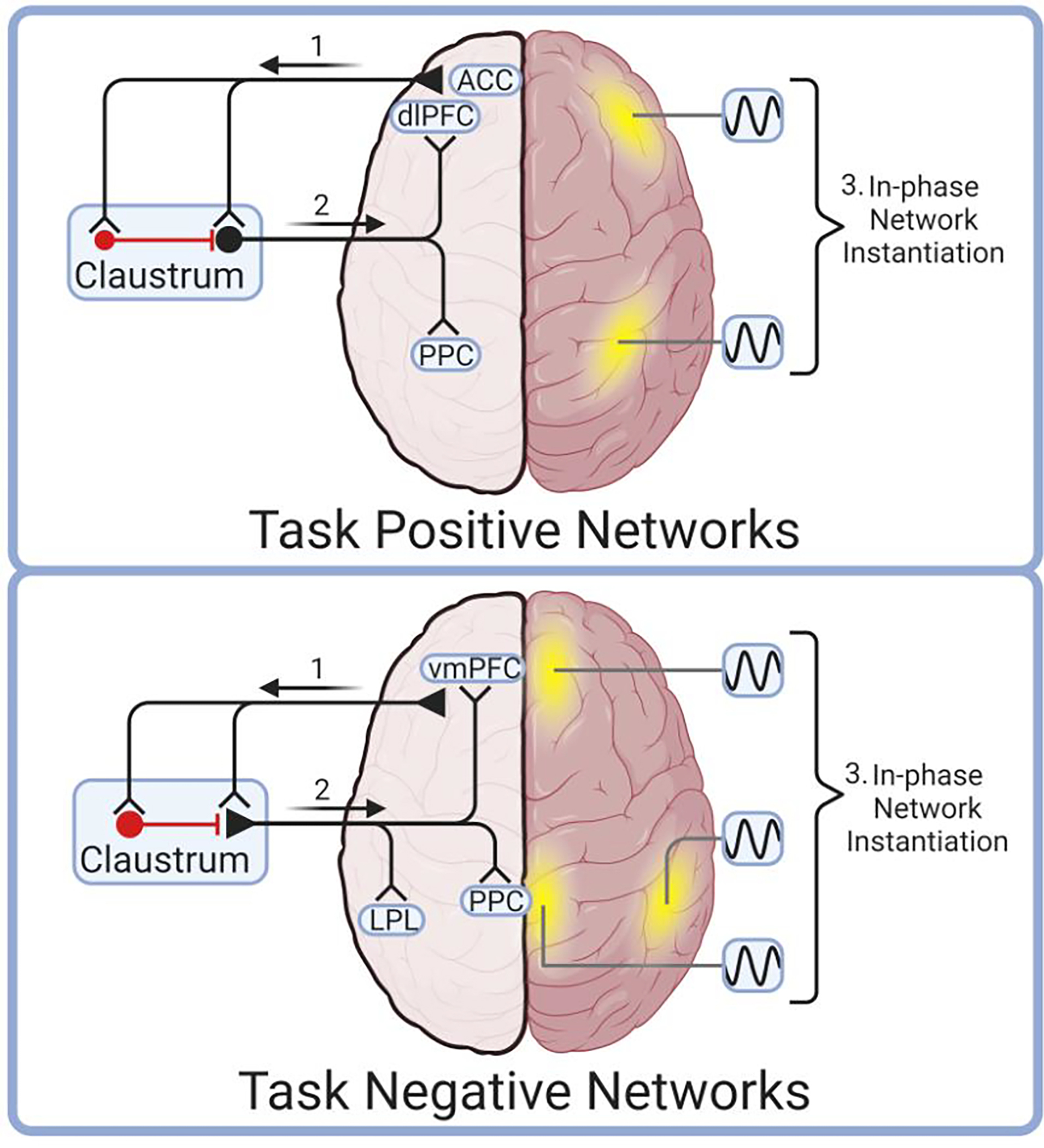
Early hypotheses of claustrum function were fueled by neuroanatomical data and yielded suggestions that the claustrum is involved in processes ranging from salience detection to multisensory integration for perceptual binding. While these hypotheses spurred useful investigations, incompatibilities inherent in these views must be reconciled to further conceptualize claustrum function amid a wealth of new data. Here, we review the varied models of claustrum function and synthesize them with developments in the field to produce a novel functional model: network instantiation in cognitive control (NICC). This model proposes that frontal cortices direct the claustrum to flexibly instantiate cortical networks to subserve cognitive control. We present literature support for this model and provide testable predictions arising from this conceptual framework.

Keywords: attention; cognition; cognitive network; cortex; cortical network; working memory.


Conflict of interest statement

Declaration of interests F.S.B. is on the scientific advisory board for Wavepaths, Ltd. and a scientific advisor for Mindstate Design Labs, Inc.

Figures

Figure 1.
Figure 1.. The location of the claustrum in mouse, monkey, and human.
The claustrum (red, CL) in mouse lies lateral to the striatum (Str) and external capsule of white matter and is positioned within the deep layers of insular cortex (IC) [145]. The claustrum in primates is located between two white matter (WM) structures, the external and extreme capsules. Lateral to the primate extreme capsule of white matter lies the insular cortex (IC), and medial to the external capsule of white matter are the striatal caudate (Cd) and putamen (Pu). The claustrum is likely present in all mammals [–16], in at least some reptiles [17], and an analogous structure is identified in avian species [19]. As such, the claustrum is speculated to have formed early in the evolution of amniotes [146,147]. This figure was created using BioRender.
Figure 2.
Figure 2.. Anatomical connections of the claustrum in rodents, cats, and monkeys.
A survey of known inputs to the claustrum (left) and outputs (right) from the claustrum. Cortical (top) and subcortical (bottom) connections are depicted. Studies used to generate this figure are reviewed in Table S1. Abbreviations: A1, primary auditory cortex; ACC, anterior cingulate cortex; AP, anterior pole; IPL, inferior parietal lobule; M1, primary motor cortex; PtA, parietal association cortex; S1, primary somatosensory cortex; V1/V2, primary and secondary visual cortex.
Figure 3.
Figure 3.. The Network Instantiation in Cognitive Control (NICC) model.
The NICC model proposes that frontal regions send cognitive control information to claustrum (1), which then transforms, amplifies, and broadcasts this information to cortical network nodes (2). These cortical nodes are brought into phase by claustro-cortical input to instantiate task-positive (top) or task-negative (bottom) cortical networks (3). In this model, unique frontal cortical inputs project to unique claustrum projection neurons that selectively target, and therefore instantiate, a given network composed of cortical nodes (yellow). For instance, the anterior cingulate cortex (ACC) is proposed to activate claustrum neurons that selectively synchronize components of the task positive network, such as the dorsolateral prefrontal cortex (dlPFC) and posterior parietal cortex (PPC) (top). Other networks may be instantiated in a similar fashion by frontal cortical activation of claustrum, which synchronizes, for instance, task negative network nodes including the ventromedial prefrontal cortex (vmPFC), posterior cingulate (PCC) and the lateral parietal lobe (LPL). This figure was created using BioRender.
Figure 4.
Figure 4.. The claustrum is functionally connected with many cortical networks at rest.
Human cortical networks are listed, as defined by the Power Atlas [66], along with network component regions, the associated functions of each network, and the functional connectivity of the claustrum with each network as previously determined [113]. Red and blue bars represent left and right claustrum functional connectivity, respectively. Abbreviations: aPFC, anterior prefrontal cortex; aIPS, anterior intraparietal sulcus; CeA, central amygdala; C-O, cingulo-opercular; Ctx, cortex; dACC, dorsal anterior cingulate cortex; dlPFC, dorsolateral prefrontal cortex; FEF, frontal eye field; F-P, frontoparietal; IFJ, inferior frontal junction; IPL, inferior parietal lobe; IPS, intraparietal sulcus; M1, primary motor cortex; mPFC, medial prefrontal cortex; MT, middle temporal visual area; OFC, orbitofrontal cortex; PCC, posterior cingulate cortex; pIPS, posterior intraparietal sulcus; S1, primary somatosensory cortex; SPL, superior parietal lobule; SSM, sensory/somatomotor; V3a; visual cortex (V3 accessory cortex);.
Figure 5.
Figure 5.. Cortico-claustro-cortical processing.
Claustrum projection neurons receive strong input from cortex, specifically frontal regions [11,95,97] (A), and are strongly inhibited by an interconnected local parvalbumin-positive interneuron population (red). Claustrum projection neurons course predominantly to cortex with weaker innervation of neighboring claustrum interneurons [124]; a subset of claustrum projection neurons burst fire in response to activation (Type II), while others exhibit only single action potential responses (Type I) [22]. Together, this configuration of claustrum microcircuitry allows for sufficient cortical input (that which exceeds the inhibitory “threshold” imposed by local interneurons) to be transformed and amplified for claustro-cortical broadcast (B). This figure was created using BioRender.
Figure 6.
Figure 6.. Claustrum innervation and modulation of cortex.
Claustrum afferents are biased toward deeper layers of cortex and to neuropeptide Y (NPY) and parvalbumin (PV)-expressing interneurons. The exception to this is a lack of claustro-cortical input to layer V parietal cortex in mouse [97], which likely reflects a lack of claustrum control of layer V neurons projecting to dorsolateral striatum/putamen. Task-positive cortical network components (e.g., prefrontal cortex and parietal cortex) at cognitive rest display relatively desynchronized oscillations (A). Claustrum firing initially elicits strong inhibition of pyramidal neurons via activation of PV-expressing and NPY-expressing interneurons, followed by resumption of pyramidal neuron excitation mediated, in part, by direct claustro-cortical input (B). This induces a phase-locking of activity across cortical regions, from which subsequent oscillatory frequencies may be governed by cortico-cortical and thalamocortical circuits. With disparate cortical regions now participating in phase-locked activity, a synchronized network is instantiated (C). This figure was created using BioRender.

Similar articles

Cited by

References

    1. Macchi G et al. (1981) The organization of the claustroneocortical projections in the cat studied by means of the HRP retrograde axonal transport. J. Comp. Neurol. 195, 681–695. 10.1002/cne.901950411 - DOI - PubMed
    1. Baizer JS et al. (1997) Projections from the claustrum to the prelunate gyrus in the monkey. Exp. Brain Res. 113, 564–568. 10.1007/pl00005607 - DOI - PubMed
    1. Sadowski M et al. (1997) Rat’s claustrum shows two main cortico-related zones. Brain Res. 756, 147–152 - PubMed
    1. Tanne-Gariepy J et al. (2002) Projections of the claustrum to the primary motor, premotor, and prefrontal cortices in the macaque monkey. J. Comp. Neurol. 454, 140–157. 10.1002/cne.10425 - DOI - PubMed
    1. Hur EE and Zaborszky L (2005) Vglut2 afferents to the medial prefrontal and primary somatosensory cortices: a combined retrograde tracing in situ hybridization study [corrected]. J. Comp. Neurol. 483, 351–373. 10.1002/cne.20444 - DOI - PubMed

Publication types