. 2022 Dec;227(9):2897-2908.
doi: 10.1007/s00429-022-02575-x. Epub 2022 Oct 3.

Stroke disconnectome decodes reading networks

Affiliations

Stroke disconnectome decodes reading networks

Stephanie J Forkel et al. Brain Struct Funct. 2022 Dec.

Abstract

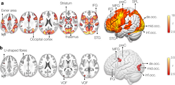
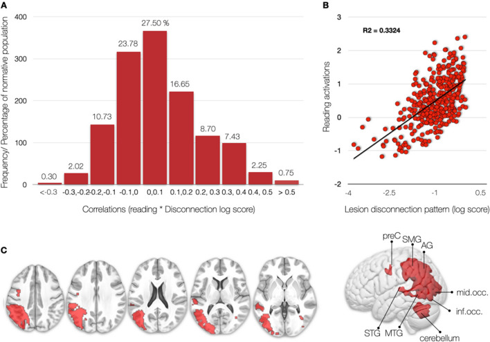
Cognitive functional neuroimaging has been around for over 30 years and has shed light on the brain areas relevant for reading. However, new methodological developments enable mapping the interaction between functional imaging and the underlying white matter networks. In this study, we used such a novel method, called the disconnectome, to decode the reading circuitry in the brain. We used the resulting disconnection patterns to predict a typical lesion that would lead to reading deficits after brain damage. Our results suggest that white matter connections critical for reading include fronto-parietal U-shaped fibres and the vertical occipital fasciculus (VOF). The lesion most predictive of a reading deficit would impinge on the left temporal, occipital, and inferior parietal gyri. This novel framework can systematically be applied to bridge the gap between the neuropathology of language and cognitive neuroscience.

Keywords: Disconnection; Exner; Language; Reading; Stroke; VOF; fMRI.


Conflict of interest statement

The authors declare no conflict of interest.

Figures

Fig. 1
Fig. 1
Schematic overview of the processing steps implemented in this study
Fig. 2
Fig. 2
Schematic description of the functional MRI paradigm for word-list reading. An event was initiated by presenting a scrambled picture for one second, followed by the presentation of a list of words (days, months, seasons) that participants were instructed to read. Upon task completion, participants responded with a button press. Subsequently, they had to indicate when the central cross changed into a square by clicking the button. (Figure amended from Hesling et al. 2019)
Fig. 3
Fig. 3
Multidimensionality of the reading network. A Average bilateral activation during the reading task (fMRI). B Locations where disconnection components explained activations during the reading task. t: The t values indicate the size of the difference relative to the variation in the condition (in panel A) and the strength of the relationship between loadings (i.e. extracted from the linear regression between components and activation patterns) and each voxel of the components maps (in panel B). In both cases, the greater the magnitude of t, the greater the evidence against the null hypothesis. IFG inferior frontal gyrus, MFG middle frontal gyrus, preC precentral gyrus, SPL superior parietal lobe, STG superior temporal gyrus, MTG middle temporal gyrus, de.occ descending occipital gyrus, mid.occ middle occipital gyrus, inf.occ inferior occipital gyrus, SMG supramarginal gyrus, AG angular gyrus, VOF vertical occipital fasciculus
Fig. 4
Fig. 4
Distribution of reading activation correlations with disconnection pattern maps in 1333 patients. A According to our experimental framework, a “pure lesion” to the reading network would only occur in 0.75% of cases. By pure lesion, we mean lesions whose disconnection profile correlates with the reading pattern of activation with a large effect size (r > 0.5). B An example of a large effect size is the correlation between the pattern of functional reading task activations and the pattern of brain disconnection of a single patient. Each dot corresponds to a brain region. This disconnection profile explains 33% of the variance functional reading task activations. C Display of the lesion pattern of the “pure lesion” to the reading network

Similar articles

Cited by

References

    1. Abdolalizadeh A, Mohammadi S, Aarabi MH. The forgotten tract of vision in multiple sclerosis: vertical occipital fasciculus, its fiber properties, and visuospatial memory. Brain Struct Funct. 2022;227(4):1479–1490. - PubMed
    1. Alario F-X, Chainay H, Lehericy S, Cohen L. The role of the supplementary motor area (SMA) in word production. Brain Res. 2006;1076:129–143. - PubMed
    1. Anderson SW, Damasio AR, Damasio H. Troubled letters but not numbers. Domain specific cognitive impairments following focal damage in frontal cortex. Brain. 1990;113(Pt 3):749–766. - PubMed
    1. Baldo JV, Kacinik N, Ludy C, Paulraj S, Moncrief A, Piai V, Curran B, Turken A, Herron T, Dronkers NF. Voxel-based lesion analysis of brain regions underlying reading and writing. Neuropsychologia. 2018;115:51–59. - PMC - PubMed
    1. Binder JR, Desai RH, Graves WW, Conant LL. Where is the semantic system? A critical review and meta-analysis of 120 functional neuroimaging studies. Cereb Cortex. 2009;19:2767–2796. - PMC - PubMed