

Neck-shaft angle measurement in children: accuracy of the conventional radiography-based (2D) methods compared to 3D reconstructions
- PMID: 36192625
- PMCID: PMC9529964
- DOI: 10.1038/s41598-022-20832-1
Neck-shaft angle measurement in children: accuracy of the conventional radiography-based (2D) methods compared to 3D reconstructions
Erratum in
-
Author Correction: Neck-shaft angle measurement in children: accuracy of the conventional radiography-based (2D) methods compared to 3D reconstructions.Sci Rep. 2022 Dec 27;12(1):22441. doi: 10.1038/s41598-022-26238-3. Sci Rep. 2022. PMID: 36575202 Free PMC article. No abstract available.
Abstract
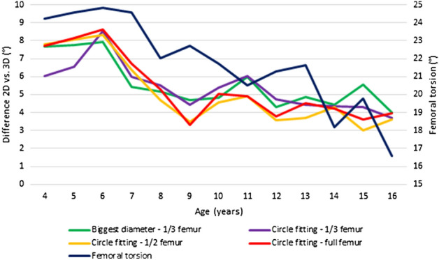
Aim of this study was to examine the accuracy of widely used conventional radiography-based (2D) neck-shaft angle measurements compared to 3D reconstruction. In our retrospective study, EOS 2D/3D images of 156 patients (312 limbs) were selected from our database (4-16 years old: 6 girls and 6 boys/year), where no pathology was revealed. Using the 2D modality of the EOS method neck-shaft angle was measured using the "biggest diameter" and "circle fitting" techniques to define the femoral neck axis and 1/3, 1/2 and full femur to determine the femoral shaft axis. EOS 3D reconstructions of same images were also performed and a comparison of 2D and 3D results was made. We did not find any significant difference between accuracy of the four examined 2D methods, although the deviation between 2 and 3D results was considerable (average difference: 5.11-5.58°, p < 0,001). In 31% of the cases, difference was more than 10°. Only femoral torsion showed significant influence on the difference (correlation coefficient: 0.380, p < 0.001). We did not find a clinically significant difference between the examined 2D methods, although their accuracy was highly questionable compared to 3D results. We suggest using any 3D imaging method for surgical planning and in uncertain cases.
© 2022. The Author(s).
Conflict of interest statement
The authors declare no competing interests.
Figures


References
-
- Grunert S, Brückl R, Rosemeyer B. [Rippstein and Müller roentgenologic determination of the actual femoral neck-shaft and antetorsion angle. 1: Correction of the conversion table and study of the effects of positioning errors] Radiologe. 1986;26:293–304. - PubMed
Publication types
MeSH terms
LinkOut - more resources
Full Text Sources
