. 2022 Oct;24(10):1541-1557.
doi: 10.1038/s41556-022-00994-w. Epub 2022 Oct 3.

Alternative RNA splicing modulates ribosomal composition and determines the spatial phenotype of glioblastoma cells

Affiliations

Alternative RNA splicing modulates ribosomal composition and determines the spatial phenotype of glioblastoma cells

Tatyana D Larionova et al. Nat Cell Biol. 2022 Oct.

Abstract

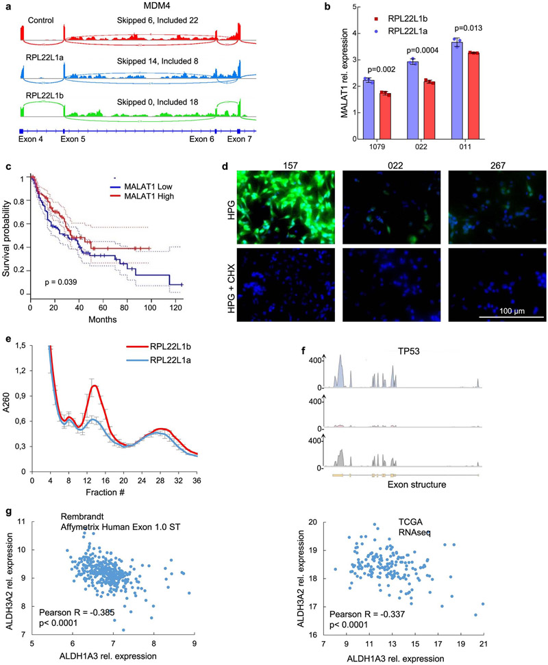
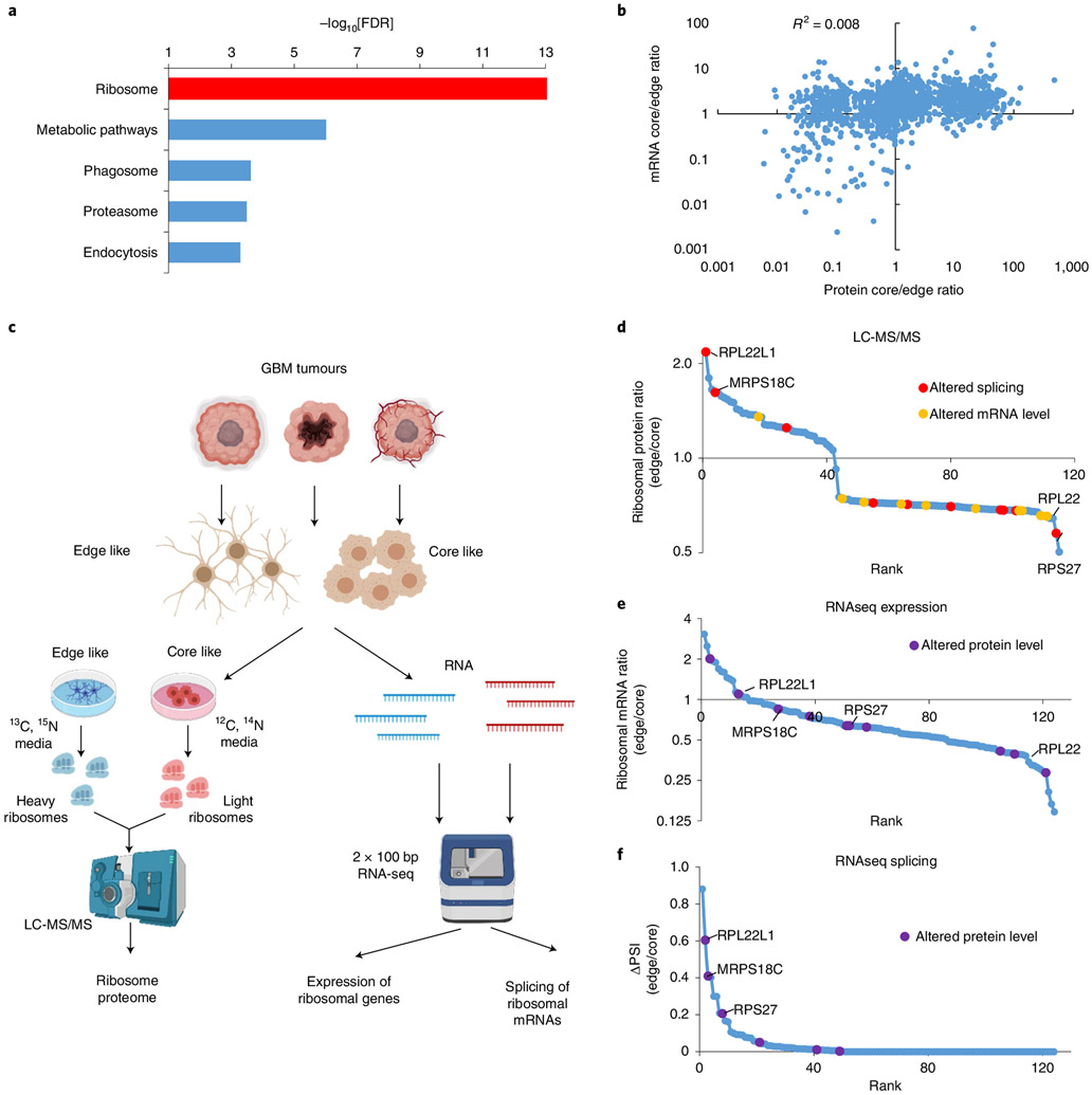
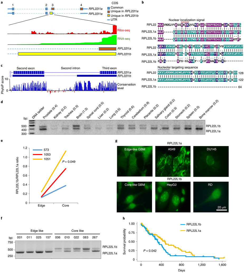
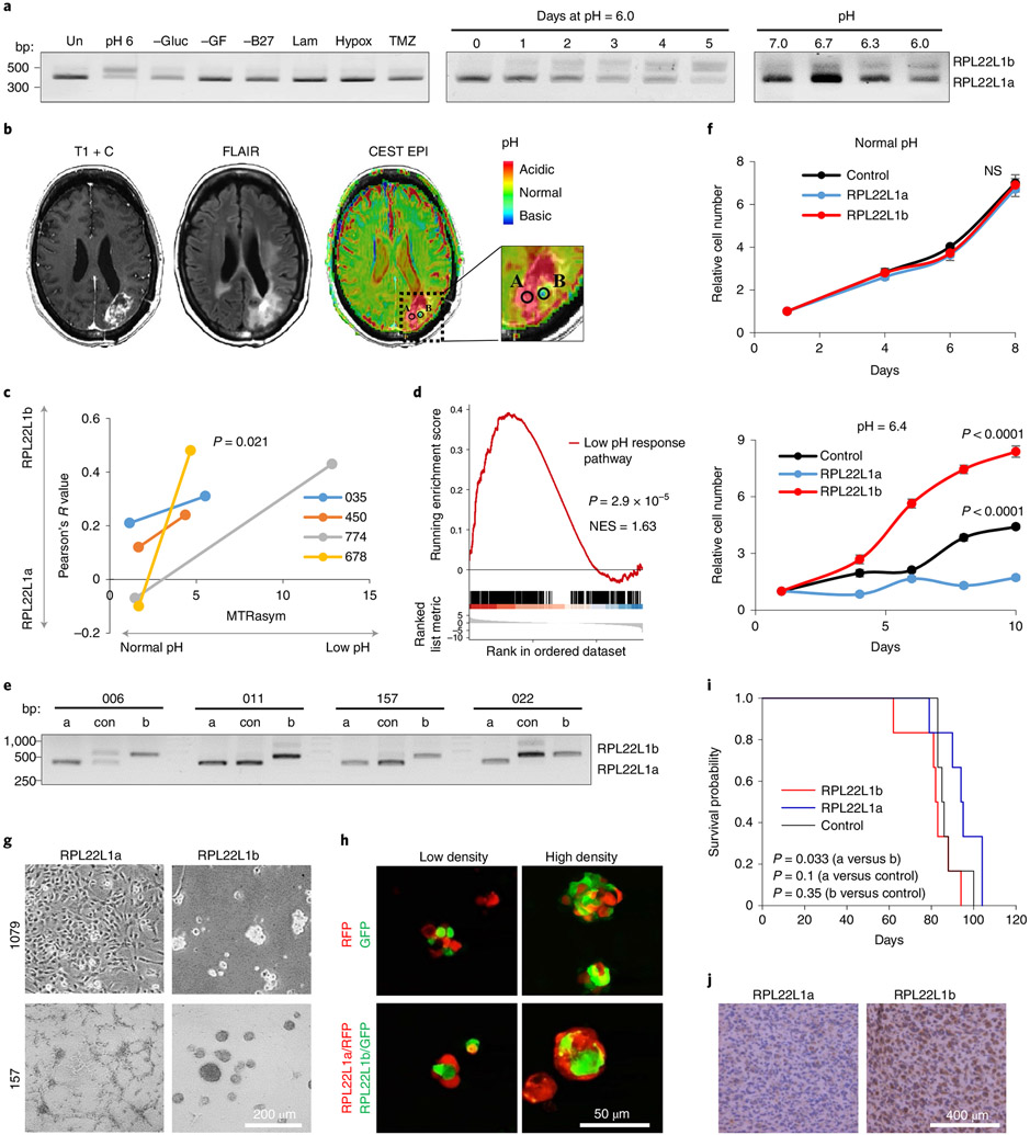
Glioblastoma (GBM) is characterized by exceptionally high intratumoral heterogeneity. However, the molecular mechanisms underlying the origin of different GBM cell populations remain unclear. Here, we found that the compositions of ribosomes of GBM cells in the tumour core and edge differ due to alternative RNA splicing. The acidic pH in the core switches before messenger RNA splicing of the ribosomal gene RPL22L1 towards the RPL22L1b isoform. This allows cells to survive acidosis, increases stemness and correlates with worse patient outcome. Mechanistically, RPL22L1b promotes RNA splicing by interacting with lncMALAT1 in the nucleus and inducing its degradation. Contrarily, in the tumour edge region, RPL22L1a interacts with ribosomes in the cytoplasm and upregulates the translation of multiple messenger RNAs including TP53. We found that the RPL22L1 isoform switch is regulated by SRSF4 and identified a compound that inhibits this process and decreases tumour growth. These findings demonstrate how distinct GBM cell populations arise during tumour growth. Targeting this mechanism may decrease GBM heterogeneity and facilitate therapy.


Figures

Extended Data Fig. 1 ∣
Extended Data Fig. 1 ∣. Proteomic and transcriptomic intratumoral heterogeneity of glioblastoma.
a, The principal component analysis of LC-MS/MS (left panel) and RNAseq (right panels) data obtained from GBM sphere lines derived from core (n = 4 different clones) and edge (n = 3 different clones) of the 1051 tumor. b, Correlation analysis of protein-to-RNA abundance in GBM sphere lines as in ‘a’. c, Correlation of protein composition of ribosomes with mRNA levels of the corresponding ribosomal genes. d, Correlation of protein composition of ribosomes with the differences in pre-mRNA splicing of the corresponding ribosomal genes. Proteins that are differentially included into ribosomes of GBM sphere lines with edge and core phenotype were determined by SILAC LC-MS/MS. Differences in mRNA levels and in pre-mRNA splicing were determined by RNA sequencing of the corresponding cells. Correlation coefficient, trend line and the most differentially present proteins/mRNAs are indicated. e, RT-PCR analysis RPL22L1 splicing in GBM cells from n = 3 different patients. PCR products were separated using PAAG electrophoresis. f, Sashimi plots demonstrating differences in splicing of RPL22L1 between GBM sphere lines with edge (157, 011, 025) and core (083, 028, 006) phenotype (cells isolated from n = 6 different patients). g, RT-PCR analysis of RPL22L1 splicing in different human cell lines. h, The principal component analysis of RNAseq data obtained from GBM sphere lines used in this study. Red – previously characterized GBM spheres with core phenotype, blue - previously characterized GBM spheres with edge phenotype, gray – sphere lines established in this study (n = 14 different patients). i, RT-PCR analysis of RPL22L1 isoform abundance in 267 cells cultivated for 5 days in normal (pH 7.4) or acidified (pH 6.0) medium. NMD inhibitor (NMDI-14) was added to a final concentration of 5 μM 24 hours before cells were collected for RNA purification.
Extended Data Fig. 2 ∣
Extended Data Fig. 2 ∣. Expression of RPL22L1 isoforms in human cancers.
a, Relative expression of RPL22L1 isoforms and SETD4 (previously described NMD target36) in Hela cells depleted for different NMD factors (experiment was performed in n = 3 biological replicates; data are mean ± SD). Raw RNAseq data were obtained from GSE152437. b, Riboseq and RNAseq read densities for RPL22L1 in U251 GBM cells (data were obtained from GSE141013 dataset). c, Polysome profile of 083 GBM spheres (upper panel) and RT-PCR analysis of RPL22L1 splicing (lower panel). d, Mass spectrometry identification of the peptide related to RPL22L1b isoform (data were obtained from PXD004023 dataset). e, Western blotting analysis of GBM spheres with edge (157, 025) and core (022, 083) phenotypes with different antibodies against RPL22L1.
Extended Data Fig. 3 ∣
Extended Data Fig. 3 ∣. Representative immunofluorescent staining of GBM cells predominantly expressing RPL22L1a (001, 157, 025) or RPL22L1b (006, 022, 083) isoform with antibodies against N-terminal part of RPL22L1.
The red dotted line indicates areas of DAPI staining (borders of the nucleus).
Extended Data Fig. 4 ∣
Extended Data Fig. 4 ∣. Prognostic value of RPL22L1 isoforms.
a, Kaplan-Meier curve showing the overall survival of Kidney Renal Clear Cell Carcinoma (n = 532 different patients), Adrenocortical carcinoma (n = 79 different patients) and Uveal Melanoma (n = 80 different patients) patients subdivided based on the splicing of RPL22L1 (log-rank test). RNAseq data were obtained from TCGA database. b, Splicing of RPL22L1 in GBM patients with wild type and mutated IDH1 (left panel); in patients younger and older than 50 years (middle panel) and in patients with high and low level of MGMT promoter methylation (right panel). 14 different probes were used to assess methylation status, none of them showed statistically significant differences in RPL22L1 splicing. Date were obtained from TCGA database (n = 154 different patients; the line in the box is the median, the up and low of the box are the first and third quartiles, and the whiskers extend to 10th and 90th percentiles respectively). Higher psi values indicate higher percentage of RPL22L1b isoform. c, FACS analysis of cell cycle distribution of 267 cells stained with propidium iodide. Populations that were collected by cell sorting are indicated. d, qRT-PCR analysis of Ki67 expression in cell populations collected as in ‘c’. e, RT-PCR analysis of RPL22L1 splicing in cell populations collected as in ‘c’ (experiment was performed in n = 3 biological replicates). All quantitative data are mean ± SD, n.s. – non significant.
Extended Data Fig. 5 ∣
Extended Data Fig. 5 ∣. RPL22L1b facilitates GBM growth in low pH conditions.
a, RT-PCR analysis of RPL22L1 splicing in 001, 011 and 022 GBM spheres that were cultivated for 5 days in normal (pH 7.4) or acidified (pH 6.0) medium. b, Immunofluorescent staining of different areas of GBM tumor tissues from patient 1051 for RPL22L1 (green) and DNA (blue). Yellow and red arrows indicate cells with nuclear and cytoplasmic localization of RPL22L1 respectively. c, Colocalization analysis of RPL22L1 and DAPI staining for the same tumor areas as in ‘b’. Pearson’s R value is indicated. d, Representative immunofluorescent staining of 022 GBM cells overexpressing RPL22L1a, RPL22L1b or an empty vector with antibodies against N-terminal part of RPL22L1. e, FACS analysis of caspase 3/7 activity and SYTOX staining of 157 glioma spheres that were transduced with lentiviruses encoding RPL22L1a, RPL22L1b or an empty vector (control) and cultivated in normal (pH 7.4) or acidified medium (pH 6.4) for 8 days.
Extended Data Fig. 6 ∣
Extended Data Fig. 6 ∣. Differential splicing of RPL22L1 promotes GBM intratumoral heterogeneity.
a, Forward vs. side scatter plot of 020 GBM spheres overexpressing RPL22L1a or RPL22L1b. Cell size was determined using Countess II Automated Cell Counter (experiment was performed in n = 4 biological replicates). b, Representative confocal images of 157 glioma spheres that were first transduced with lentiviruses encoding RPL22L1a or RPL22L1b and subsequently transduced with lentiviruses encoding GFP or RFP. Next cells overexpressing RPL22L1a + RFP were mixed with cells overexpressing RPL22L1b + GFP and imaged 4 days later. c, Representative images of wound healing assay with 157 cells stably expressing RPL22L1a, RPL22L1b or an empty vector. d, Representative IHC staining for CD109 of mouse brain sections obtained 3 months after intracranial injection of 3·105 luciferase labeled 1051 glioma spheres overexpressing RPL22L1a or RPL22L1b (n = 2 mice per group). e, PAAG electrophoresis of recombinant RPL22L1a (a), or RPL22L1b (b), that were purified from E. coli. f, Enrichment analysis of proteins that were bound to recombinant His-tagged RPL22L1a (upper panel) or RPL22L1b (lower panel) and subsequently identified by LC-MS/MS.
Extended Data Fig. 7 ∣
Extended Data Fig. 7 ∣. Molecular functions RPL22L1 isoforms in GBM cells.
a, Enrichment analysis of proteins that were co-purified with Fc-tagged RPL22L1a (lower panel) or RPL22L1b (upper panel) and subsequently identified by LC-MS/MS. b, Enrichment analysis of mRNAs that were co-purified with Fc-tagged RPL22L1a (left panel) or RPL22L1b (right panel) and subsequently identified by RNA sequencing. c, KEGG database GSVA analysis of RNA sequencing data obtained from 157 cells overexpressing RPL22L1a, RPL22L1b or an empty vector (experiment was performed in two biological replicates). d, Enrichment analysis of proteins that were differentially present in 157 cells overexpressing RPL22L1a or RPL22L1b isoform as determined by SILAC LC-MS/MS. e, Enrichment analysis of alternative splicing events related to exon skipping (upper panel) or mutually exclusive exon inclusion (lover panel) detected in 157 cells stably expressing RPL22L1b compared to the control cells.
Extended Data Fig. 8 ∣
Extended Data Fig. 8 ∣. RPL22L1 isoforms regulate pre-mRNA splicing and mRNA translation in GBM cells.
a, Sashimi plots demonstrating differences in splicing of MDM4 between 157 GBM spheres overexpressing RPL22L1a, RPL22L1b or an empty vector. Number of reeds that confirms exon skipping or exon inclusion is indicated. b, qRT-PCR analysis of MALAT1 RNA stability in 1079, 022 and 011 GBM spheres overexpressing RPL22L1a or RPL22L1b isoforms. Cells were cultivated for 6 hours with Actinomycin D at final concentration of 10 μg/ml (experiment was performed in n = 3 biological replicates). c, Kaplan-Meier curve showing the disease-free survival of glioma patients subdivided based on the MALAT1 expression level (n = 338 different patients, log-rank test). Data were obtained from TCGA database. d, Representative fluorescence images demonstrating L-homopropargylglycine (HPG) incorporation into newly synthesized proteins in 157, 022 and 267 GBM sphere lines. HPG was detected by Alexa Fluor488 azide (green). Nucleus were visualized by DAPI (blue). Cells pretreated for 30 min with cycloheximide (100 μg/ml) were used as a control. e, Polysome profiles of 1079 GBM spheres stably expressing RPL22L1a or RPL22L1b (experiment was performed in n = 2 biological replicates). f, RNA-IP enrichment profiles of RPL22L1a, RPL22L1b and a control protein for TP53 gene. 157 cells overexpressing Fc-tagged proteins were used for the experiment. g, Correlation of ALDH3A2 and ALDH1A3 expression levels in glioma. Data were obtained from REMBRANDT (n = 354 different patients; left panel) or TCGA (n = 671 different patients; right panel) databases. All quantitative data are mean ± SD.
Extended Data Fig. 9 ∣
Extended Data Fig. 9 ∣. SRSF4 regulates RPL22L1 splicing.
a, SRSF4 CLIP-tag enrichment profile in Rpl22l1 RNA sequence. Data were obtained from E-MTAB-747 dataset. b, qRT-PCR analysis of RNAs that were co-purified with Fc-tagged SRSF4 (red), or a control protein (blue) with primers for RPL22L1 exon 3, RPL22L1 3’UTR or RPL22 (experiment was performed in n = 3 biological replicates). c, SRSF4 expression level in different regions of GBM tumor (n = 122 RNA samples obtained from n = 10 different patients, one-way ANOVA test, following Dunnett’s/Tukey’s posttest). Data were obtained from IVY GAP database. The line in the box is the median, the up and low of the box are the first and third quartiles, and the whiskers extend to 10th and 90th percentiles respectively. d, qRT-PCR analysis of SRSF4 expression in 006, 030 and 157 GBM cells transduced with lentiviruses encoding non-target shRNA (shNT) or two different shRNAs against SRSF4 (48shSRSF4 and 49shSRSF4). e, Quantification of RPL22L1 splicing differences in 030 (upper) and 157 (lower) GBM cells transduced with lentiviruses as in ‘d’ (experiment was performed in n = 3 biological replicates). Higher psi values indicate higher percentage of RPL22L1b isoform. f, Kaplan-Meier curve showing the overall survival of glioblastoma patients (n = 179) subdivided based on the SRSF4 expression level (log-rank test). Data were obtained from REMBRANDT database. g, RT-PCR analysis of RPL22L1 splicing in 011 and 1079 glioma spheres that were left untreated (Un) or treated for 24 hours with CMP3a (10 μM), EY404 (10 μM), Fg1059 (10 μM), Cisplatin (10 μM), TMZ (100 μM) or Pladienolide B (1 μM). h, Enrichment analysis of the proteins that were differentially phosphorylated (more than 10 fold differences) in 157 GBM cells after 3 and 12 hours of incubation with 3 μM of FG1059 as opposed to untreated cells (experiment was performed in n = 2 biological replicates). Reactom and InterPro databases were used to calculate enrichment. All quantitative data are mean ± SD.
Extended Data Fig. 10 ∣
Extended Data Fig. 10 ∣. FG1059 induce apoptosis of GBM cells.
a, In vitro cell viability assay of 022, 157 and 1079 GBM spheres stably expressing RPL22L1a, RPL22L1b or an empty vector. Cells were treated with different concentrations of FG1059 for 5 days (experiment was performed in n = 6 biological replicates; data are mean ± SD). b, FACS analysis of caspase 3/7 activity and SYTOX staining of 157 cells treated with DMSO; 0.2 μM of FG1059; 20 μM of TMZ or with both compounds simultaneously for 24 hours. c, FACS analysis of caspase 3/7 activity and SYTOX staining of 157 cells treated with DMSO; 50 nM of FG1059; 50 nM of Pladienolide B or with both compounds simultaneously for 24 hours. d, Kaplan-Meier curve showing the overall survival of glioma (n = 509 different patients; left panel) and glioblastoma (n = 152 different patients; right panel) patients subdivided based on the RPL22 expression level (log-rank test). Data were obtained from TCGA database. e, Flow cytometry gating used for apoptosis assay (left panel) and for CD133 or HPG staining (right panel).
Fig. 1 ∣
Fig. 1 ∣. GBM cells from the edge and core of the tumour have ribosomes with different protein compositions.
a, Kyoto Encyclopedia of Genes and Genomes enrichment analysis of proteins differentially present (fold change > 4; spectral count > 2) between the edge and core of the tumour. FDR, false discovery rate. b, Correlation between the proteome and transcriptome of edge and core GBM tissue. c, Experimental workflow used to study differences in the protein composition of ribosomes. bp, base pair. d, Relative levels of ribosomal proteins identified by SILAC LC-MS/MS in ribosomes purified from GBM sphere lines with edge versus core phenotypes. e, Relative mRNA levels of ribosomal genes in GBM sphere lines with edge versus core phenotypes, as identified by RNA-seq. f, Differences in the splicing of ribosomal genes between GBM sphere lines with edge and core phenotypes, as identified by RNA-seq. PSI, percentage spliced in index1. In d–f, the top ten differentially present proteins, differentially expressed mRNAs and differentially spliced pre-mRNAs are highlighted in violet, yellow and red, respectively. RNA-seq was performed for neurospheres obtained from n = 6 different patients.
Fig. 2 ∣
Fig. 2 ∣. Alternative splicing generates two different isoforms of RPL22L1.
a, Schematic of RPL22L1 isoforms together with ribo-seq and RNA-seq read densities aligned to the corresponding regions of the genome. The data were obtained from GWIPs-viz Browser. The first, second, third and fourth exons are indicated by the numbers 1–4 at the top. CDS, coding sequence; UTR, untranslated region. b, Alignment of the amino acid sequences of RPL22, RPL22L1a and RPL22L1b (shadings indicate positions which have a single fully conserved residue in all three proteins (purple); in any two proteins (green) or residue with similar properties in two proteins (blue); number of amino acids in the protein sequence is indicated). c, Pattern of genome conservation among 100 vertebrates for the RPL22L1 region spanning from the second to the third exon. d, RT-PCR analysis of RPL22L1 splicing in different human tissues. Approximate RPL22L1b/RPL22L1a isoform ratios are indicated. e, Ratios of RPL22L1 isoforms in paired GBM tissue isolated from edge and core regions of tumours (tissue from n = 3 different patients; Wilcoxon signed rank-sum test). The data were obtained from RNA-seq. f, RT-PCR analysis of RPL22L1 splicing in GBM sphere lines with edge (001, 011, 025 and 157) and core (006, 010, 022, 083 and 267) phenotypes (spheres isolated from n = 9 different patients). g, Representative immunofluorescence stainings of cells predominantly expressing the RPL22L1a isoform (GBM spheres 157, A549 and DU145) or RPL22L1b isoform (GBM spheres 267, HepG2 and RD) with antibodies against the N-terminal part of RPL22L1. h, Kaplan–Meier curve showing the overall survival of patients with GBM, subdivided into two groups based on the splicing of RPL22L1 (n = 154 different patients; log-rank test). The data were obtained from the TCGA database.
Fig. 3 ∣
Fig. 3 ∣. The RPL22L1 isoform ratio is regulated by extracellular pH.
a, RT-PCR analysis of RPL22L1 splicing in 157 GBM spheres cultivated under different conditions for the indicated periods of time. Un, untreated cells in control medium; pH 6, acidified medium; −Gluc, without glucose; −GF, without growth factors; −B27, without nutrition supplement; Lam, cultured as a monolayer on laminin; Hypox, cultured under hypoxia; TMZ, cultured in the presence of 50 μM temozolomide (similar data were obtained for cells isolated from n = 4 different patients; see Extended Data Fig. 3a). b, Representative contrast-enhanced T1-weighted (left), fluid attenuated inversion recovery (FLAIR; middle) and pH–weighted amine CEST–EPI (MTRasym at 3 ppm; right) MRI images of GBM tumours. Biopsy regions with low and normal pH are labelled A and B, respectively. c, Colocalization coefficient of RPL22L1 and DAPI staining plotted against MTRasym values. MTRasym represents the acidity of the tumour region. The colocalization coefficient reflects the RPL22L1b/RPL22L1a isoform ratio (samples were obtained from n = 4 different patients; Wilcoxon signed rank-sum test). d, GSEA of RNA-seq data from RPL22Lb/RPL22L1ahigh versus RPL22Lb/RPL22L1alow GBM tissue. The data were obtained from the TCGA database (n = 77 different patients). NES, normalized enrichment score. e, RT-PCR analysis of RPL22L1 splicing in GBM cells transduced with lentiviruses encoding RPL22L1a (a), RPL22L1b (b) or an empty vector (con) (n = 4 different patients). f, In vitro cell growth assay of 157 glioma spheres overexpressing RPL22L1a, RPL22L1b or an empty vector (control) and cultivated in normal (pH 7.4; top) or acidified medium (pH 6.4; bottom) (the experiment was performed in n = 6 biological replicates; unpaired two-tailed t-test). NS, not significant. g, Representative microscopic images of 1079 and 157 glioma spheres overexpressing RPL22L1a or RPL22L1b (similar data were obtained for cells isolated from n = 4 different patients). h, Representative fluorescence images of 1079 glioma spheres overexpressing GFP, RFP, RFP+ RPL22L1a or GFP+ RPL22L1b. The cells were mixed in different densities and imaged 24 h later (similar data were obtained for cells isolated from n = 2 different patients). i, Kaplan–Meier survival curves of mice injected with 3 × 10⁵ luciferase-labelled 1051 glioma spheres overexpressing RPL22L1a, RPL22L1b or an empty vector (n = 6 mice per group; log-rank test). j, Representative immunohistochemical staining for CD109 of brain sections from mice injected as in i (n = 2 mice per group; see Extended Data Fig. 6d). All quantitative data are means ± s.d.
Fig. 4 ∣
Fig. 4 ∣. Interactome of RPL22L1 isoforms.
a, Venn diagram representing proteins that were bound to recombinant His-tagged RPL22L1a (blue), RPL22L1b (red) or control beads (green) and subsequently identified by LC-MS/MS. Proteins that were detected only in RPL22L1a or only in RPL22L1b samples were subjected to enrichment analysis. The most significantly enriched terms and corresponding P values are indicated. b, Venn diagram representing proteins that were co-purified with Fc-tagged RPL22L1a (blue), RPL22L1b (red) or control protein (green) and subsequently identified by LC-MS/MS. Proteins that were present in the RPL22L1a sample or RPL22L1b sample and absent in the control sample were subjected to enrichment analysis. The most significantly enriched terms and corresponding P values are indicated. KEGG, Kyoto Encyclopedia of Genes and Genomes. c, Capillary electrophoresis of RNA that was co-purified with Fc-tagged RPL22L1a or RPL22L1b. d, Venn diagram representing RNAs that were co-purified with Fc-tagged RPL22L1a (blue), RPL22L1b (red) or control protein (green) and subsequently identified by RNA-seq. RNA was considered differentially present if the fold change was >6 and the number of transcripts per million (tpm) was >0.5. Differentially present RNAs were subjected to enrichment analysis. The most significantly enriched terms and corresponding P values are indicated. e, Hallmark database GSVA of RNA-seq data obtained from 157 cells overexpressing RPL22L1a, RPL22L1b or an empty vector (sequencing was performed in n = 2 biological replicates). IFN-α, interferon-α; NF-κB, nuclear factor-κB; TNF-α, tumour necrosis factor-α; TGF-β, transforming growth factor-β. f, Results of SILAC LC-MS/MS analysis of 157 cells overexpressing RPL22L1a (grown in heavy isotope medium) or RPL22L1b (grown in regular isotope medium). Proteins were considered differentially expressed if the difference between the intensity-based absolute quantification value for heavy and light peptides was >50%. Differentially present proteins were subjected to KEGG enrichment analysis. The graph indicates the ratio of proteins (in RPL22L1b versus RPL22L1a samples) related to the most significantly enriched clusters (the experiment was performed in n = 2 biological replicates).
Fig. 5 ∣
Fig. 5 ∣. Molecular functions of RPL22L1b.
a, Pie chart representing the number and type of alternative splicing events detected in 157 cells expressing RPL22L1b compared with control cells. b, Volcano plot showing significantly different splicing events related to exon skipping (left) and intron retention (right) detected in samples as in a (sequencing was performed in n = 2 biological replicates). c, qRT-PCR analysis of the MDM4 isoform expression ratios between 157 GBM spheres overexpressing RPL22L1a, RPL22L1b or an empty vector (the experiment was performed in n = 3 biological replicates; unpaired two-tailed t-test). d, RNA-IP enrichment profiles of RPL22L1a, RPL22L1b and a control protein for MALAT1. 157 cells overexpressing Fc-tagged proteins were used for the experiment. e, qRT-PCR analysis of samples as in d with primers for MALAT1 (the experiment was performed in n = 3 biological replicates; unpaired two-tailed t-test). f, qRT-PCR analysis of MALAT1 expression in 157 cells stably expressing different isoforms of RPL22L1 (the experiment was performed in n = 3 biological replicates). g, Analysis of MALAT1 RNA stability at different time points after actinomycin D treatment (10 μg ml−1) of cells as in f (similar data were obtained for cells isolated from n = 4 different patients; see Extended Data Fig. 7b; for each sphere line, the experiment was performed in n = 3 biological replicates; unpaired two-tailed t-test). h, Kaplan–Meier curve showing the overall survival of patients with glioma, subdivided based on the MALAT1 expression level (n = 338 different patients; log-rank test; the data were obtained from the TCGA database). i, FACS analysis for CD133 staining of GBM spheres expressing RPL22L1 isoforms (the cells were isolated from n = 2 different patients). j, qRT-PCR analysis of ALDH1A3, CD133, Nanog, Oct4, Sox2 and GFAP expression in 157 cells stably overexpressing RPL22L1a, RPL22L1b or an empty vector (the experiment was performed in n = 3 biological replicates; unpaired two-tailed t-test). k, Neurosphere formation assay of cells expressing RPL22L1a or RPL22L1b (the cells were isolated from n = 3 different patients). The line within each box represents the mean stem cell frequency. The top and bottom edges of each box are the upper and lower estimated confidence intervals for stem cell frequency. All quantitative data are means ± s.d.
Fig. 6 ∣
Fig. 6 ∣. Molecular functions of RPL22L1a.
a, FACS analysis of HPG incorporation into newly synthesized proteins in 022 cells overexpressing RPL22L1a, RPL22L1b or an empty vector. Cells pre-treated for 30 min with cycloheximide (100 μg ml−1) were used as a negative control. b, FACS analysis of HPG incorporation into newly synthesized proteins in 001, 025 and 157 GBM sphere lines (predominantly expressing RPL22L1a) and 006, 010 and 022 GBM sphere lines (predominantly expressing RPL22L1b) (the cells were isolated from n = 6 different patients). c, Polysome profiles of 1079 GBM spheres stably expressing RPL22L1a or RPL22L1b (similar data were obtained for cells isolated from n = 2 different patients; see Extended Data Fig. 7e). d, RNA-IP enrichment profiles of RPL22L1a, RPL22L1b and a control protein for the genes CDK5, ALDH3A2 and RPN2. 157 cells overexpressing Fc-tagged proteins were used for the experiment. e, qRT-PCR analysis of RNA-IP samples as in c with primers for TP53, CDK5, ALDH3A2 and RPN2 (the experiment was performed in n = 3 biological replicates; unpaired two-tailed t-test). f, qRT-PCR analysis of TP53, CDK5, ALDH3A2 and RPN2 expression in 157 cells expressing the different isoforms of RPL22L1 (the experiment was performed in n = 3 biological replicates). g, Western blotting analysis of GBM spheres overexpressing RPL22L1a, RPL22L1b or an empty vector (the cells were isolated from n = 3 different patients). The numbers below the panels indicate the approximate band intensity, which was first normalized to the actin β level and then to the intensity of the band in the corresponding control sample. All quantitative data are means ± s.d.
Fig. 7 ∣
Fig. 7 ∣. SRSF4 regulates the splicing of RPL22L1.
a, Correlation between the RPL22L1b/RPL22L1a isoform ratio and the expression levels of different splicing factors. The x axis shows the correlation coefficient for all cancer samples (n = 7,631 different patients). The y axis represents the correlation for GBM samples (n = 154 different patients). The data were obtained from the TCGA database. b, SRSF4 mRNA levels in paired edge and core GBM samples (tissue from n = 3 different patients; Wilcoxon signed rank-sum test). The data were obtained by RNA-seq. FPKM, fragments per kilobase of transcript per million mapped reads. c, Representative immunohistofluorescence staining with antibodies against SRSF4 (red) and DAPI (blue) of low- and normal-pH GBM tumour biopsy samples obtained using pH–weighted molecular MRI (from n = 2 different patients). d, qRT-PCR analysis of SRSF4 expression in 157 cells cultivated in acidified medium (pH 6.0) for 0–5 d (the experiment was performed in n = 3 biological replicates; unpaired two-tailed t-test). e, RPL22L1 isoform ratio in U87MG cells transfected with plasmids encoding GFP (control) or GFP-tagged SRSF1, SRSF2, SRSF3 or SRSF4 proteins and subsequently sorted for GFP using FACS (the experiment was performed in n = 3 biological replicates). f, RT-PCR analysis of RPL22L1 splicing in 157 glioma spheres overexpressing GFP or GFP-SRSF4. g, Representative microscopic images of 006 (left) and 030 (right) GBM cells transduced with lentiviruses encoding non-target small hairpin RNA (shNT) or small hairpin RNAs against SRSF4 (shSRSF4). The cells were attached to the laminin-coated surface. h, RT-PCR analysis of RPL22L1 splicing in 006, 030 and 157 GBM cells transduced with lentiviruses encoding shNT or two different small hairpin RNAs against SRSF4 (48shSRSF4 and 49shSRSF4). i, qRT-PCR analysis of CD133 and ALDH1A3 expression in 030 (top) and 157 (bottom) GBM spheres transduced with lentiviruses as in h (the experiment was performed in n = 3 biological replicates; unpaired two-tailed t-test). j, In vitro cell growth assay of cells as in i (the experiment was performed in n = 6 biological replicates; unpaired two-tailed t-test). k, Kaplan–Meier curve showing the overall survival of patients with glioma (n = 446) subdivided based on the SRSF4 expression level (log-rank test). The data were obtained from the REMBRANDT database. All quantitative data are means±s.d.
Fig. 8 ∣
Fig. 8 ∣. FG1059 impairs the splicing of RPL22L1.
a, RT-PCR analysis of RPL22L1 splicing in 157 glioma spheres that were left untreated (Un) or treated for 24 h with CMP3a, EY404 (EY), FG1059 (FG) or pladienolide B (Pl-B) (similar data were obtained for cells isolated from n = 3 different patients; see Extended Data Fig. 8f). b, Chemical structure of FG1059. c, Results of Sanger sequencing of the RPL22L1 isoform purified from FG1059-treated cells. d, Principal component analysis of the phosphoproteome of 157 GBM cells treated with 3 μM FG1059 for 0, 3, 6 or 12 h. e, Representative fluorescence images of 157 cells transfected with a plasmid encoding RFP-SRSF4 and subsequently treated with 5 μM FG1059 for 18 h. The DNA was stained with DAPI. DMSO, dimethyl sulfoxide. f, In vitro cell viability assay of GBM spheres obtained from n = 7 different patients and normal human astrocytes (NHAs) that were treated with various concentrations of FG1059 for 5 d (the experiment was performed in n = 6 biological replicates). Cells predominantly expressing the RPL22L1a (001, 025 and 157) and RPL22L1b (006, 022, 030 and 267) isoforms are indicated. g, Representative immunohistochemical staining of GBM intracranial xerographs for Ki-67 and ALDH1A3. Immunocompromised mice were injected with 5 × 10⁵ patient-derived 1763 glioma cells and treated 1 month later with FG1059 (10 mg kg−1 tail vein injection five times every 3 d). Mice were sacrificed 3 d after the last injections to obtain brain slices (n = 5 mice in the control group and n = 3 mice in the FG1059-treated group). h, Quantification of immunohistochemical staining for Ki-67 in the samples as in g, using the German immunohistochemical scoring (GIS) system (unpaired two-tailed t-test). i, Same as in h, but for ALDH1A3 staining (unpaired two-tailed t-test). j, Top, experimental workflow used to study the effect of FG1059 on tumour growth in vivo. Bottom, Kaplan–Meier survival curves of mice intracranially injected with 5 × 10⁵ 1763 glioma cells and subsequently intraperitoneally injected with FG1059 or a solvent (n = 6 and n = 8 mice per treatment and control group, respectively; log-rank test). k, Proposed molecular mechanism of GBM spatial phenotype regulation by RPL22L1 isoforms. All quantitative data are means ± s.d.

Comment in

  • Ribosome specialization in glioblastoma.
    Genuth NR, Barna M. Genuth NR, et al. Nat Cell Biol. 2022 Oct;24(10):1451-1453. doi: 10.1038/s41556-022-01000-z. Nat Cell Biol. 2022. PMID: 36192633 No abstract available.

References

    1. Van Linde ME et al. Treatment outcome of patients with recurrent glioblastoma multiforme: a retrospective multicenter analysis. J. Neurooncol 135, 183–192 (2017). - PMC - PubMed
    1. Puchalski RB et al. An anatomic transcriptional atlas of human glioblastoma. Science 360, 660–663 (2018). - PMC - PubMed
    1. Bastola S et al. Glioma-initiating cells at tumor edge gain signals from tumor core cells to promote their malignancy. Nat. Commun 11, 4660 (2020). - PMC - PubMed
    1. Hjelmeland AB et al. Acidic stress promotes a glioma stem cell phenotype. Cell Death Differ. 18, 829–840 (2011). - PMC - PubMed
    1. Vaupel P, Kallinowski F & Okunieff P Blood flow, oxygen and nutrient supply, and metabolic microenvironment of human tumors: a review. Cancer Res. 49, 6449–6465 (1989). - PubMed

Publication types