Review
. 2022 Dec;31(12):e4463.
doi: 10.1002/pro.4463.

An evolutionary history of the CoA-binding protein Nat/Ivy

Affiliations
Review

An evolutionary history of the CoA-binding protein Nat/Ivy

Liam M Longo et al. Protein Sci. 2022 Dec.

Abstract

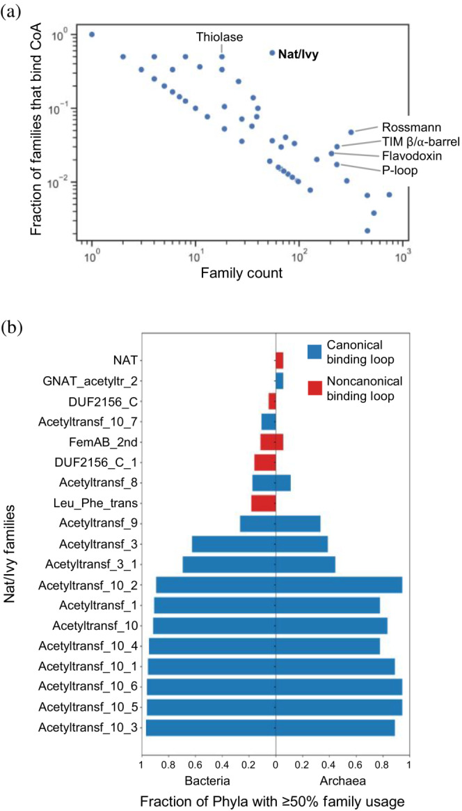
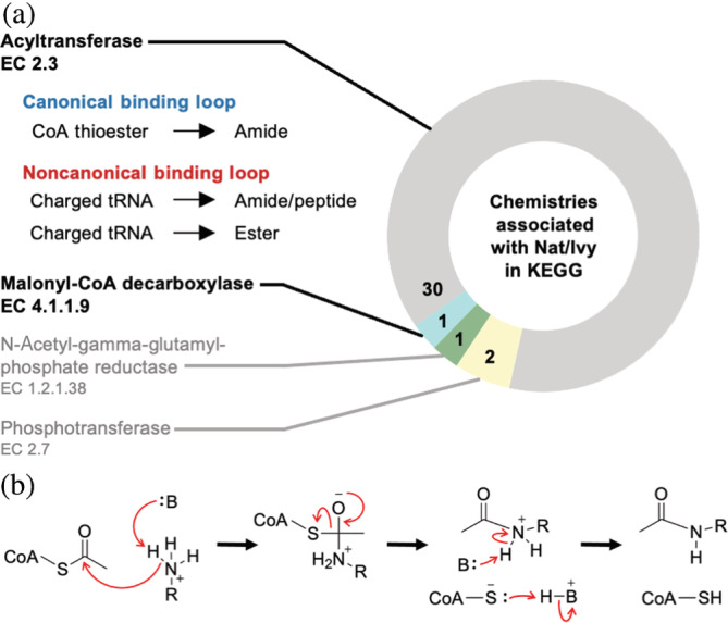
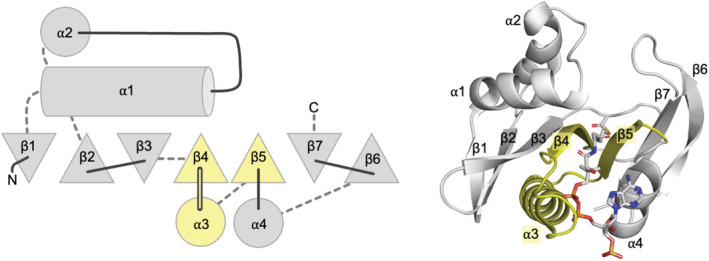
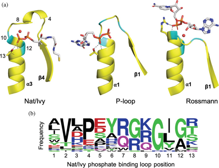
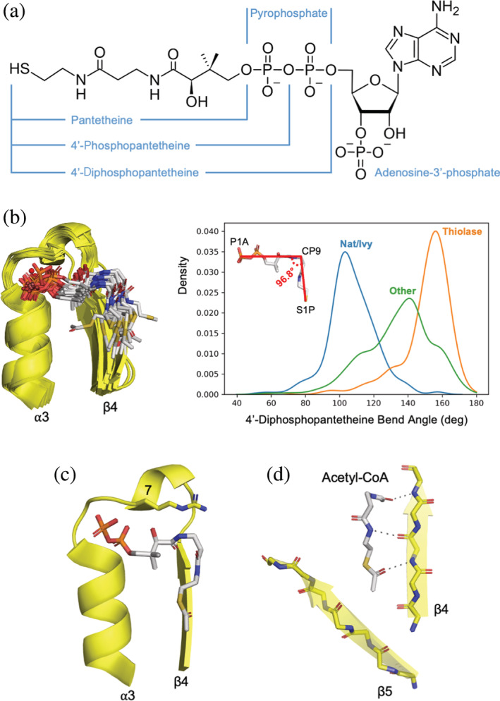
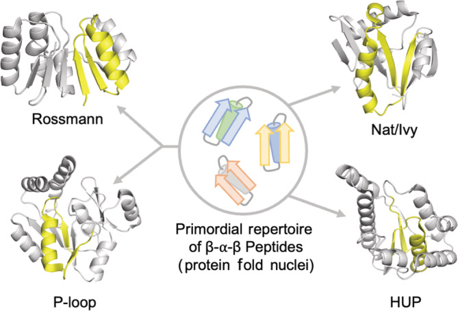
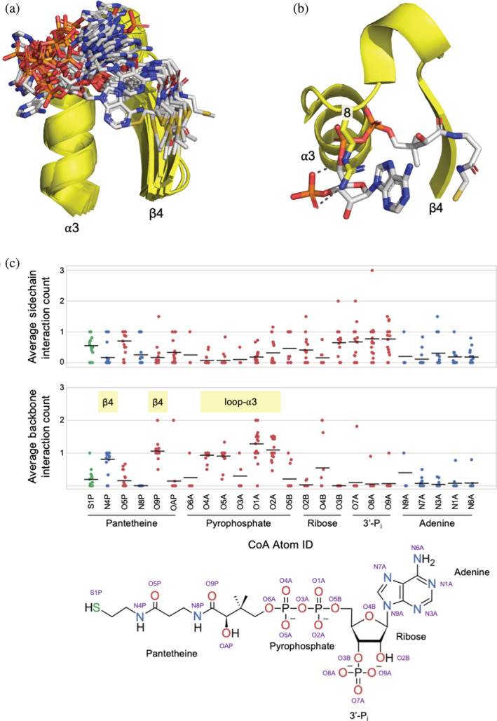
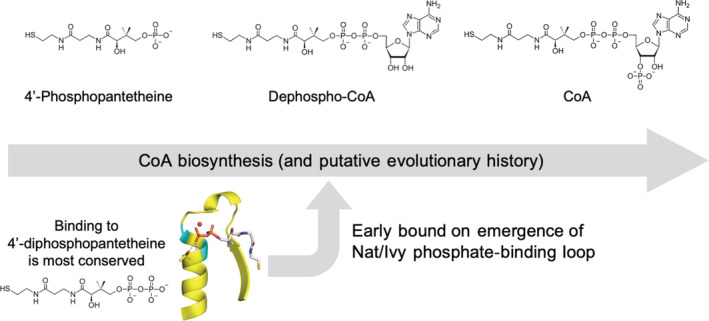
Nat/Ivy is a diverse and ubiquitous CoA-binding evolutionary lineage that catalyzes acyltransferase reactions, primarily converting thioesters into amides. At the heart of the Nat/Ivy fold is a phosphate-binding loop that bears a striking resemblance to that of P-loop NTPases-both are extended, glycine-rich loops situated between a β-strand and an α-helix. Nat/Ivy, therefore, represents an intriguing intersection between thioester chemistry, a putative primitive energy currency, and an ancient mode of phospho-ligand binding. Current evidence suggests that Nat/Ivy emerged independently of other cofactor-utilizing enzymes, and that the observed structural similarity-particularly of the cofactor binding site-is the product of shared constraints instead of shared ancestry. The reliance of Nat/Ivy on a β-α-β motif for CoA-binding highlights the extent to which this simple structural motif may have been a fundamental evolutionary "nucleus" around which modern cofactor-binding domains condensed, as has been suggested for HUP domains, Rossmanns, and P-loop NTPases. Finally, by dissecting the patterns of conserved interactions between Nat/Ivy families and CoA, the coevolution of the enzyme and the cofactor was analyzed. As with the Rossmann, it appears that the pyrophosphate moiety at the center of the cofactor predates the enzyme, suggesting that Nat/Ivy emerged sometime after the metabolite dephospho-CoA.

Keywords: GNAT; P-loop; Rossmann; acetyltransferase; coenzyme A; nucleotide cofactor; phosphate binding loop.


Figures

FIGURE 1
FIGURE 1
Nat/Ivy is a fundamental CoA‐binding domain. (a) Fraction of families (i.e., ECOD F‐groups) with a CoA or acetyl‐CoA binding event as a function of total family count. Nat/Ivy is the most diverged protein for which >50% of families bind CoA. (b) Distribution of Nat/Ivy domains in bacteria and archaea. Nat/Ivy proteins bearing a canonical CoA‐binding loop are essentially ubiquitous among microbes
FIGURE 2
FIGURE 2
(a) Chemistry associated with Nat/Ivy domains in KEGG. Nat/Ivy primarily catalyzes acyl transfer chemistry. (b) The Nat/Ivy reaction mechanism. Although there is some debate, the curated M‐CSA database favors only mechanisms without an acyl enzyme intermediate
FIGURE 3
FIGURE 3
The conserved core of Nat/Ivy domains. β‐strands are represented as triangles; triangles pointing up denote β‐stands that are coming out of the plane of the paper (toward the reader) whereas triangles pointing down denote β‐strands that are going into the plane of the paper (away from the reader). Solid lines indicate loops at the top of the fold and dashed lines indicate loops at the bottom of the fold. Structural elements shaded yellow comprise the majority of the CoA binding site. Domain e2jddA1 pictured. All structure figures were generated in PyMOL (www.pymol.org)
FIGURE 4
FIGURE 4
The canonical phosphate‐binding loop of Nat/Ivy strongly resembles those of other ancient enzymes. (a) A comparison between the phosphate binding loops of Nat/Ivy (e5kf9A2), P‐loop (e1yrbA1), and Rossmann (e1lssA1) enzymes. Conserved glycine residues are colored cyan. Conserved water molecules are rendered as red spheres. Residues forming bidentate interactions at the N‐terminus of an α‐helix are shown as sticks, with hydrogen bonds shown as dashed grey lines. For Nat/Ivy, numbers correspond to the positions in panel b. The 3′‐phosphoadenosine moiety of CoA is omitted for clarity. (b) Frequency plot derived from the consensus sequence of each Nat/Ivy family with a canonical phosphate binding loop (figure generated using WebLogo 63 )
FIGURE 5
FIGURE 5
Binding of pantetheine by Nat/Ivy. (a) The structure of CoA. (b) Overlay of the representative structures with a canonical binding loop (see Supplemental File 1 for full list). The cofactor adopts a bent conformation. (c) A “latch residue” at position 7 of the phosphate‐binding loop lays across the top of the pantetheine moiety of the bound CoA (e4jxrB2). (d) Binding in a crevice formed by β4 and β5. Note how the cofactor extends the β‐sheet by binding the edge of β4 (e4jxrB2)
FIGURE 6
FIGURE 6
β‐α‐β peptides as nuclei for the emergence cofactor binding domains. At the heart of several ancient enzymes—including Rossmanns, P‐loops, and HUPs (the evolutionary lineage that encompasses Class I aminoacyl‐tRNA synthetases)—is a β‐α‐β motif that mediates ligand binding
FIGURE 7
FIGURE 7
Binding to adenosine and the 3′‐phosphate. (a) An overlay of the representative domains reveals that the adenine moiety and the 3′‐phosphate adopt a range of conformations (c.f. Figure 6a, showing the comparatively tight conformational ensemble of the 4′‐diphosphopantetheine moiety). (b) An Arg residue at position 8 stacking on top of adenine and forming hydrogen bonds with the 3′‐phosphate (e3dddA2). (c) Family‐normalized interaction analysis between CoA and Nat/Ivy where each point corresponds to an F‐group in the Nat/Ivy evolutionary lineage. Interactions between O, N, and S atoms (3.2 Å distance cutoff) between CoA and protein for each family with a canonical phosphate‐binding loop were identified and classified as either a sidechain or backbone interaction. Each point corresponds to a family average and the bar indicates the average of all family averages. Whereas binding to the pyrophosphate and pantetheine moieties of CoA are both associated with conserved backbone interactions (indicated with yellow bars), binding to the nucleoside and the 3′‐phosphate are not. There is no case where a sidechain interaction is completely conserved
FIGURE 8
FIGURE 8
Coevolution of the Nat/Ivy phosphate‐binding loop and the CoA cofactor. The most conserved binding interactions—and those interactions that are mediated by the protein backbone—are centered on the 4′‐diphosphopantetheine moiety of CoA. However, the biosynthesis of CoA, which may report on its evolutionary history, does not involve a stage with a free 4′‐diphosphopantetheine metabolite. Instead, 4′‐diphosphopantetheine is, to the best of current knowledge, absent from contemporary metabolism. Assuming there was no stage during which 4′‐diphosphopantetheine was the major form of CoA, the emergence of the phosphate‐binding loop of Nat/Ivy seems to have occurred after the emergence of dephospho‐CoA (see main text for a more detailed discussion). This is in keeping with the Rossmann fold, which binds the pyrophosphate moiety of the dinucleotide cofactor NAD/P and therefore seems to have emerged after the appearance of the full dinucleotide cofactor

References

    1. Krtenic B, Drazic A, Arnesen T, Reuter N. Classification and phylogeny for the annotation of novel eukaryotic GNAT acetyltransferases. PLoS Comput Biol. 2020;16:e1007988. - PMC - PubMed
    1. Kornprobst M, Turk M, Kellner N, et al. Architecture of the 90S pre‐ribosome: A structural view on the birth of the eukaryotic ribosome. Cell. 2016;166:380–393. - PubMed
    1. Dong SH, Frane ND, Christensen QH, Greenberg EP, Nagarajan R, Nair SK. Molecular basis for the substrate specificity of quorum signal synthases. Proc Natl Acad Sci U S A. 2017;114:9092–9097. - PMC - PubMed
    1. Agarwal V, Metlitskaya A, Severinov K, Nair SK. Structural basis for microcin C7 inactivation by the MccE acetyltransferase. J Biol Chem. 2011;286:21295–21303. - PMC - PubMed
    1. Oda K, Matoba Y, Noda M, Kumagai T, Sugiyama M. Catalytic mechanism of bleomycin N‐acetyltransferase proposed on the basis of its crystal structure. J Biol Chem. 2010;285:1446–1456. - PMC - PubMed

Publication types