. 2022 Sep 16;25(10):105154.
doi: 10.1016/j.isci.2022.105154. eCollection 2022 Oct 21.

Adam10-dependent Notch signaling establishes dental epithelial cell boundaries required for enamel formation

Affiliations

Adam10-dependent Notch signaling establishes dental epithelial cell boundaries required for enamel formation

Thimios A Mitsiadis et al. iScience. .

Abstract

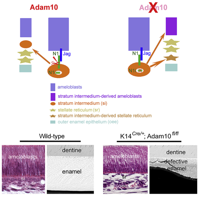
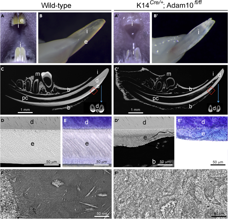
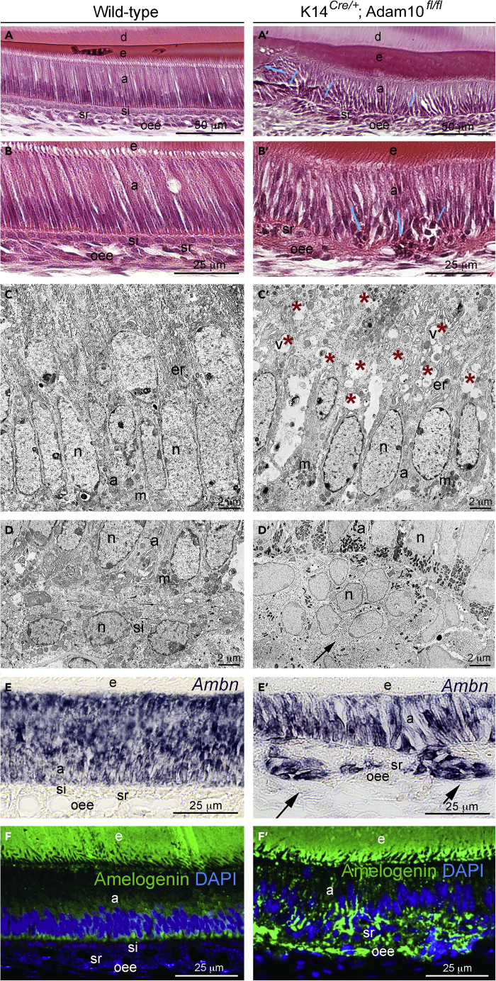
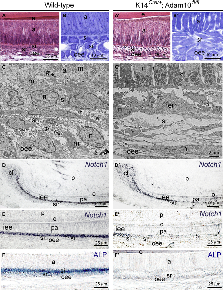
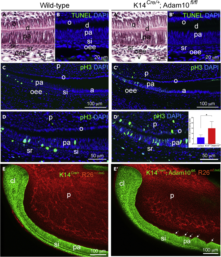
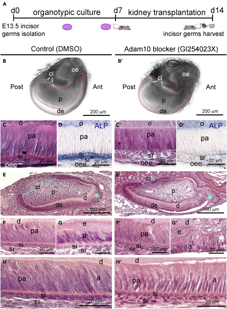
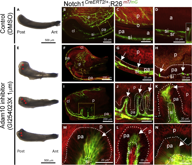
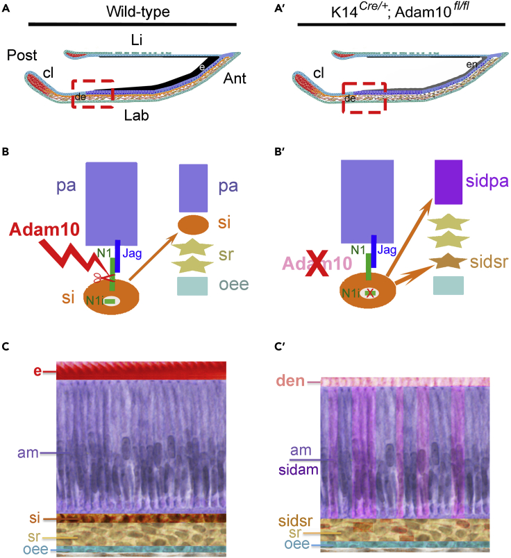
The disintegrin and metalloproteinase Adam10 is a membrane-bound sheddase that regulates Notch signaling and ensures epidermal integrity. To address the function of Adam10 in the continuously growing incisors, we used Keratin14 Cre/+;Adam10 fl/fl transgenic mice, in which Adam10 is conditionally deleted in the dental epithelium. Keratin14 Cre/+;Adam10 fl/fl mice exhibited severe abnormalities, including defective enamel formation reminiscent of human enamel pathologies. Histological analyses of mutant incisors revealed absence of stratum intermedium, and severe disorganization of enamel-secreting ameloblasts. In situ hybridization and immunostaining analyses in the Keratin14 Cre/+;Adam10 fl/fl incisors showed strong Notch1 downregulation in dental epithelium and ectopic distribution of enamel-specific molecules, including ameloblastin and amelogenin. Lineage tracing studies using Notch1 CreERT2 ;R26 mT/mG mice demonstrated that loss of the stratum intermedium cells was due to their fate switch toward the ameloblast lineage. Overall, our data reveal that in the continuously growing incisors the Adam10/Notch axis controls dental epithelial cell boundaries, cell fate switch and proper enamel formation.

Keywords: Cell biology; Developmental biology; Model organism.


Conflict of interest statement

The authors have no conflicts of interest to report.

Figures

None
Graphical abstract
Figure 1
Figure 1
Adam10 deletion in the dental epithelium causes enamel defects (A-B′) Macroscopic views of wild-type (A, B) and K14Cre/+;Adam10fl/fl (A′, B′) incisors. Mutant incisors appear white/transparent (compare A with A′, and B with B′). (C, C′) Scanning Electron Microscopy (SEM) images of lower jaws from wild-type (C) and mutant (C′) mice. Images in the lower right corner are sections from wild-type and mutant jaws processed by Microcomputed Tomography (μCT). (D, D′) High magnification SEM images of red boxed areas on the labial side of the incisors in (C) and (C′), showing normal enamel in wild-type incisors (D) and the hypoplastic and hypomineralized enamel in K14Cre/+;Adam10fl/fl incisors (D′). (E, E′) Toluidine blue staining in ground sections showing the well-structured white (absence of organic matrix) enamel in wild-type incisors (E) and deficient blue stained (high levels of organic matrix) enamel in K14Cre/+; Adam10fl/fl incisors (E′). (F, F′) High magnification transmission electron microscopy (TEM) images showing enamel crystals and intercrystallite spaces in wild-type (F) and K14Cre/+;Adam10fl/fl (F′) incisors. Notice the existence of large intercrystallite spaces in the enamel of mutant incisors (F′). Abbreviations: b, bone; d, dentine; e, enamel; i, incisor; m, molar; pc, pulp chamber.
Figure 2
Figure 2
Adam10 deletion induces disorganization of the ameloblast layer, loss of ameloblasts polarity and ectopic enamel secretion (A-B’) Hematoxylin and eosin (H&E) staining of wild-type (A, B) and K14Cre/+;Adam10fl/fl (A′, B′) incisors. Blue arrows indicate areas of ectopically formed cell clusters (A′, B′). Higher magnification H&E images showing ameloblasts orientation in wild-type (B) and K14Cre/+;Adam10fl/fl (B′) incisors. (C, C′) TEM analysis of ameloblasts from wild-type (C) and K14Cre/+;Adam10fl/fl (C′) incisors demonstrating the increased number of vacuoles (red asterisks) and empty spaces in the ameloblasts of K14Cre/+;Adam10fl/fl incisors (C′). (D, D′) TEM images of structures adjacent to ameloblast layer from C and C′ of wild-type (D) and K14Cre/+;Adam10fl/fl (D′) incisors, demonstrating intact stratum intermedium in wild-type incisors (D), and the presence of cell clusters (black arrow) in K14Cre/+;Adam10fl/fl incisors (D′). (E, E′) In situ hybridization showing expression of Ambn (violet color) in the wild-type (E) and K14Cre/+;Adam10fl/fl (E′) incisors. In K14Cre/+;Adam10fl/fl incisors, Ambn is expressed by ameloblasts and clusters of dental epithelial cells (black arrows in E′). (F, F′) Immunofluorescent staining showing distribution of the Amelogenin protein (green color) =in wild-type (F) and K14Cre/+;Adam10fl/fl (F′) incisors. Cell nuclei are identified by DAPI (blue color). Abbreviation: a, ameloblasts; d, dentine; e, enamel; er, endoplasmic reticulum; m, mitochondria; n, nucleus; oee, outer enamel epithelium; si, stratum intermedium; sr, stellate reticulum; v, vacuoles.
Figure 3
Figure 3
Adam10 deletion induces loss of the stratum intermedium, downregulation of the Notch1 expression and loss of the Alkaline Phosphatase (ALP) activity (A, A′) H&E staining of wild-type (A) and K14Cre/+;Adam10fl/fl (A′) incisors. (B, B′) High magnification images of Toluidine blue staining in wild-type (B) and K14Cre/+;Adam10fl/fl (B′) incisors. (C, C′) TEM images of the interface between ameloblasts and stratum intermedium in wild-type incisors (C) and the interface between ameloblasts and stellate reticulum cells in K14Cre/+;Adam10fl/fl incisors (C′). (D-D′) In situ hybridization showing Notch1 expression in wild-type (D) and K14Cre/+;Adam10fl/fl (D′) incisors. Notice that in K14Cre/+;Adam10fl/fl incisors, Notch1 expression is downregulated outside of the cervical loop area, soon after the onset of dental epithelial cell differentiation. (E, E′) Higher magnifications of dental epithelial cells layers from D and D′, respectively, demonstrating downregulated Notch1 expression following differentiation of inner enamel epithelium cells into preameloblasts. (F, F′) ALP staining in wild-type (F) and K14Cre/+;Adam10fl/fl (F′) incisors, from the region of differentiating ameloblasts. Abbreviations: a, ameloblasts; b, bone; cl, cervical loop; d, dentine; e, enamel; er, endoplasmic reticulum; i, incisor; iee, inner enamel epithelium; m, mitochondria; n, nucleus; o, odontoblasts; oee, outer enamel epithelium; p, dental pulp; pa, preameloblasts; si, stratum intermedium; sr, stellate reticulum.
Figure 4
Figure 4
Adam10 deletion affects cell proliferation, but not apoptotic events (A-B′) H&E (A, A′) and TUNEL (B, B′) staining in wild-type (A, B) and K14Cre/+;Adam10fl/fl (A′, B′) incisors showing absence of cell death (green cells) in K14Cre/+;Adam10fl/fl incisors (B′). DAPI staining (blue color) identifies the cell nuclei. (C-D′) Low (C, C′) and high (D, D′) magnification images of the immunofluorescent staining against Phosphorylated Histone 3 (pH3; green color) indicates higher cell proliferation activity in the preameloblast region of K14Cre/+;Adam10fl/fl incisors (C′, D′), compared to wild-type (C, D). Cell nuclei are identified by DAPI (blue color). Inserted box in (D′) shows the quantification of the number of pH3-positive cells in the preameloblast layer of the wild-type (blue color) and K14Cre/+;Adam10fl/fl (red color) incisors. Asterisk (∗) indicates the p value = 0.0176. (E, E′) Whole mount imaging showing the dental epithelium in green (mGFP) in K14Cre/+;R26mT/mG (E; control) and K14Cre/+;Adam10fl/fl;R26mT/mG (E′, mutant) incisors. White arrows in (E′) indicate uneven dental epithelium upon Adam10 deletion. mTomato in red color labels the dental pulp mesenchyme. Abbreviations: a, ameloblasts; cl, cervical loop; d, dentine; e, enamel; o, odontoblasts; oee, outer enamel epithelium; p, dental pulp; pa, preameloblasts; si, stratum intermedium; sr, stellate reticulum.
Figure 5
Figure 5
The Adam10 inhibitor GI254023X recapitulates the tooth phenotype induced by genetic deletion of Adam10 (A) Schematic representation of the experimental setup. (B, B′) Images of E13.5 incisor germs cultured for seven days under control conditions (B) or in presence of GI254023X (B′). (C-D′) High magnification images of H&E (C, C′) and ALP staining (D, D′) in control (C, D) and GI254023X-treated (C′, D′) incisors, focusing on the differentiated epithelial cell layers, and showing the loss of stratum intermedium (C′) and ALP staining (D′) in GI254023X-treated incisors. E, E′). H&E staining in control (E) and GI254023X-treated (E′) incisors after kidney capsule transplantation. Asterisks indicate the most anterior (tip) part of the incisors. Notice the disorganized dental epithelium at the anterior part of GI254023X-treated incisors (asterisk in E′). (F-H′) Higher magnifications of the H&E stained median (F, F′, H, H′) and anterior (G, G′) parts of the control (F, G, H) and GI254023X-treated (F′, G′, H′) incisors. Reestablishment of the ameloblast and stratum intermedium layers at the median part of GI254023X-treated incisors (F′, H′). Absence of stratum intermedium and ameloblasts disorganization in the anterior part of GI254023X-treated incisors (G′). Abbreviations: a, ameloblasts; Ant, anterior part; cl, cervical loop; d, dentine; de, dental epithelium; p, dental pulp; Post, posterior part; e, enamel; o, odontoblasts; oe, oral epithelium; oee, outer enamel epithelium; pa, preameloblasts; si, stratum intermedium; sr, stellate reticulum.
Figure 6
Figure 6
Lineage tracing of Notch1-expressing cells upon GI254023X Adam10 inhibition (A) Brightfield image of the Notch1CreERT2/+;R26mT/mG incisor cultured for three days in control conditions. (B–D) Confocal images showing different regions of the control incisor (A), demonstrating GFP fluorescence in the cuboidal-shaped stratum intermedium cells (B-D) and sporadically in the fully differentiated, elongated ameloblasts (insert in D). Image (C) is high magnification of the yellow dotted area of image (B). White dotted lines indicate the margins of the preameloblast layer from the dental pulp and the stratum intermedium layer. (E) Notch1CreERT2/+;R26mT/mG incisors cultured for one to four days in presence of GI25423X. Red arrows point to the folding areas within the preameloblast layer at the posterior part of the incisors. (F–H) Confocal images showing GFP fluorescence in the Notch1CreERT2/+;R26mT/mG incisors cultured for one day in presence of the inhibitor. Image (G) is high magnification of the yellow dotted area of image (F). White arrows point to GFP-positive cells in the preameloblast layer. (I–N) Confocal images indicating GFP fluorescent staining in the Notch1CreERT2/+;R26mT/mG incisors cultured for four days in presence of the inhibitor. Image (J) is high magnification of the yellow dotted area of image (I) Images (K-N) are high magnifications of the folding areas within the preameloblast layer located at the posterior part of the incisors. White arrows point to the GFP-positive preameloblasts within the folded part of the preameloblast layer. White dotted lines indicate the margins of the preameloblast layer from the dental pulp and the stratum intermedium layer. Abbreviations: a, ameloblasts; Ant, anterior part; cl, cervical loop; e, enamel; o, odontoblasts; p, dental pulp; pa, preameloblasts; Post, posterior part; si, stratum intermedium; sr, stellate reticulum.
Figure 7
Figure 7
Hypothetical model illustrating the function of Adam10 in the mouse incisor (A, A′) Schematic representation of wild-type (A) and K14Cre/+;Adam10fl/fl (A′) incisors showing the absence of stratum intermedium and the deposition of a dysfunctional enamel upon Adam10 deletion. Red dashed boxes indicate the areas where stratum intermedium cell fate change starts and the stratum intermedium cells adopt the preameloblast and stellate reticulum cell fates in the K14Cre/+;Adam10fl/fl incisors (A′). (B, B′) Schematic representation of the mechanism of Adam10 action in the dental epithelium. Adam10 is responsible for the cleavage of Notch1 in stratum intermedium cells (red scissors) that enables nuclear translocation of the Notch1 intracellular domain (B; Notch1i, in green). Depletion of Adam10 in the incisor epithelium disables nuclear translocation of the Notch1 intracellular domain, thus deregulating the function of the Notch pathway. This initiates conversion of a portion of stratum intermedium cells into preameloblasts and consequent ameloblast disorganization and impaired enamel formation. In addition, another portion of stratum intermedium cells flattens and becomes stellate reticulum cells participating in the formation of the incisor’s papillary cell layer. (C, C′) Cartoon depicting the definitive alterations in the number and composition of the epithelial cell layers (each of them represented by a specific color) and enamel formation in the K14Cre/+;Adam10fl/fl incisors. Stratum intermedium (orange color) cell fate change toward the stellate reticulum and preameloblast/ameloblast fate generates the additional stratum intermedium-derived stellate reticulum (enhanced brown color) and stratum intermedium-derived ameloblasts (enhanced violet color) and leads to defective enamel deposition. Abbreviations: am, ameloblasts; Ant, anterior part; cl, cervical loop; de, dental epithelium; den, dysfunctional enamel; e, enamel; Jag, Jagged; Lab, labial side; Li, lingual side; N1, Notch1; N1i, intracellular Notch1; oee, outer enamel epithelium; pa, preameloblasts; Post, posterior part; si, stratum intermedium; sidam, stratum intermedium-derived ameloblasts; sidpa, stratum intermedium-derived preameloblasts; sr, stellate reticulum.

Similar articles

Cited by

References

    1. Andersson E.R., Sandberg R., Lendahl U. Notch signaling: simplicity in design, versatility in function. Development. 2011;138:3593–3612. - PubMed
    1. Artavanis-Tsakonas S., Matsuno K., Fortini M.E. Notch signaling. Science. 1995;268:225–232. - PubMed
    1. Artavanis-Tsakonas S., Muskavitch M.A.T. Notch: the past, the present, and the future. Curr. Top. Dev. Biol. 2010;92:1–29. - PubMed
    1. Artavanis-Tsakonas S., Rand M.D., Lake R.J. Notch signaling: cell fate control and signal integration in development. Science. 1999;284:770–776. - PubMed
    1. Balic A., Thesleff I. Tissue interactions regulating tooth development and renewal. Curr. Top. Dev. Biol. 2015;115:157–186. - PubMed

LinkOut - more resources