Review
. 2022 Sep 16;25(10):105124.
doi: 10.1016/j.isci.2022.105124. eCollection 2022 Oct 21.

Clinical neuroscience and neurotechnology: An amazing symbiosis

Affiliations
Review

Clinical neuroscience and neurotechnology: An amazing symbiosis

Andrea Cometa et al. iScience. .

Abstract

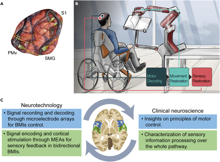
In the last decades, clinical neuroscience found a novel ally in neurotechnologies, devices able to record and stimulate electrical activity in the nervous system. These technologies improved the ability to diagnose and treat neural disorders. Neurotechnologies are concurrently enabling a deeper understanding of healthy and pathological dynamics of the nervous system through stimulation and recordings during brain implants. On the other hand, clinical neurosciences are not only driving neuroengineering toward the most relevant clinical issues, but are also shaping the neurotechnologies thanks to clinical advancements. For instance, understanding the etiology of a disease informs the location of a therapeutic stimulation, but also the way stimulation patterns should be designed to be more effective/naturalistic. Here, we describe cases of fruitful integration such as Deep Brain Stimulation and cortical interfaces to highlight how this symbiosis between clinical neuroscience and neurotechnology is closer to a novel integrated framework than to a simple interdisciplinary interaction.

Keywords: Bioelectronics; Clinical neuroscience.


Conflict of interest statement

S.M. holds shares in the companies IUVO, GTX, and SensArs Neurotechnologies, which are all developing neurotechnologies to restore sensorimotor functions of people with disabilities. All other authors declare no competing interests.

Figures

None
Graphical abstract
Figure 1
Figure 1
Interplay between clinical neuroscience and neurotechnology Representative scheme of the positive feedback loop between neurotechnology and clinical neuroscience.
Figure 2
Figure 2
Deep-brain stimulation and basal ganglia neuroscience (A) Reconstruction of bilateral directional deep brain stimulation (DBS) electrodes (model Boston Scientific Cartesia) implanted to the subthalamic nucleus (orange), view from right posterior. The right electrode shows a stimulation volume (red) steering the current toward the target nucleus. Small white arrows surrounding the right electrode depict the magnitude and direction of the electric field generated by a 3mA anodal stimulation. Internal (green) and external (blue) pallidum and subthalamic nucleus (STN) defined by the DISTAL atlas (Ewert et al., 2018). The backdrop shows a section of the 100um postmortem brain template (Edlow et al., 2019). (B) Schematic illustration of the volume of tissue activated by a DBS electrode in the STN (putative functional divisions are represented). Active contacts (displayed in white) generate electric fields of decaying intensity (high in red, medium in orange, low in yellow and threshold in dashed black). Eddy currents that diffuse from the target regions are represented as radial black arrows. From (Vissani et al., 2020). (C) Positive feedback loop between DBS neurotechnologies (left) and associated advancements in the understanding of neural underpinnings of motor control and cognitive brain disorders (right). LFPs: local field potentials. Pink areas in the inset identifies basal ganglia.
Figure 3
Figure 3
Brain-machine interfaces and neuroscience of sensorimotor functions (A) 96-channels microelectrode arrays (MEAs) implanted into ventral premotor cortex (PMv), and supramarginal gyrus (SMG), and two 48-channel stimulating arrays implanted into primary somatosensory cortex (S1). Reproduced with permission from (Armenta Salas et al., 2018). (B) Schematic representation of bidirectional brain-machine interface (BMI) allowing user to voluntarily control external devices, such as a robotic arm. Sensors embedded in the actuator can provide sensory information to the subjects by stimulating their cortex. From (Shokur et al., 2021). (C) Clinical applications of MEAs for BMIs (left) and associated advancements in the understanding of motor control and sensory processing (right) inside the positive feedback loop. Green: somatosensory cortices; blue: motor cortices.
Figure 4
Figure 4
Intracortical neural interfaces, neurosurgery and cognitive neuroscience (A) Subdural grid versus depth electrode placement in previously operated brain tumor patients. (left) electrocorticographic (ECoG) grid. (right) Stereo-electroencephalographic (SEEG) electrodes. Reproduced with permission from (Sweet et al., 2013). (B) Intraoperative functional mapping done by analyzing the ECoG signal while the patient executes cognitive tasks during awake surgery. From (Erez et al., 2021). (C) Clinical applications of SEEG and ECoG (left) and associated advancements in the understanding of cognitive brain functions (right) inside the positive feedback loop. Red: temporal lobes.

References

    1. Al-Fatly B., Ewert S., Kübler D., Kroneberg D., Horn A., Kühn A.A. Connectivity profile of thalamic deep brain stimulation to effectively treat essential tremor. Brain. 2019;142:3086–3098. doi: 10.1093/brain/awz236. - DOI - PubMed
    1. Aman J.E., Johnson L.A., Sanabria D.E., Wang J., Patriat R., Hill M., Marshall E., MacKinnon C.D., Cooper S.E., Schrock L.E., et al. Directional deep brain stimulation leads reveal spatially distinct oscillatory activity in the globus pallidus internus of Parkinson’s disease patients. Neurobiol. Dis. 2020;139:104819. doi: 10.1016/j.nbd.2020.104819. - DOI - PMC - PubMed
    1. Anderson D.N., Charlebois C.M., Smith E.H., Arain A.M., Davis T.S., Rolston J.D. Probabilistic comparison of gray and white matter coverage between depth and surface intracranial electrodes in epilepsy. Sci. Rep. 2021;11:24155. doi: 10.1038/s41598-021-03414-5. - DOI - PMC - PubMed
    1. Anumanchipalli G.K., Chartier J., Chang E.F. Speech synthesis from neural decoding of spoken sentences. Nature. 2019;568:493–498. doi: 10.1038/s41586-019-1119-1. - DOI - PMC - PubMed
    1. Arber S., Costa R.M. Connecting neuronal circuits for movement. Science. 2018;360:1403–1404. doi: 10.1126/science.aat5994. - DOI - PubMed

LinkOut - more resources