. 2022 Sep 20:2022:9518592.
doi: 10.1155/2022/9518592. eCollection 2022.

Epidermal Growth Factor Receptor Inhibition Is Protective in Hyperoxia-Induced Lung Injury

Affiliations

Epidermal Growth Factor Receptor Inhibition Is Protective in Hyperoxia-Induced Lung Injury

Zachary M Harris et al. Oxid Med Cell Longev. .

Abstract

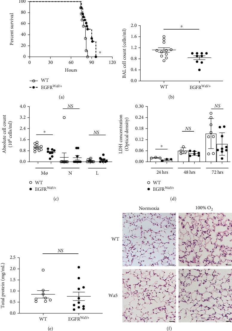
Aims: Studies have linked severe hyperoxia, or prolonged exposure to very high oxygen levels, with worse clinical outcomes. This study investigated the role of epidermal growth factor receptor (EGFR) in hyperoxia-induced lung injury at very high oxygen levels (>95%).

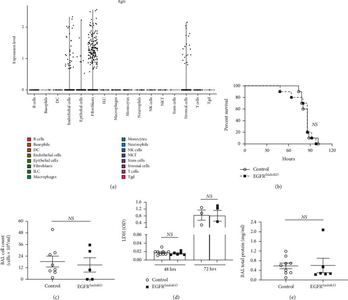
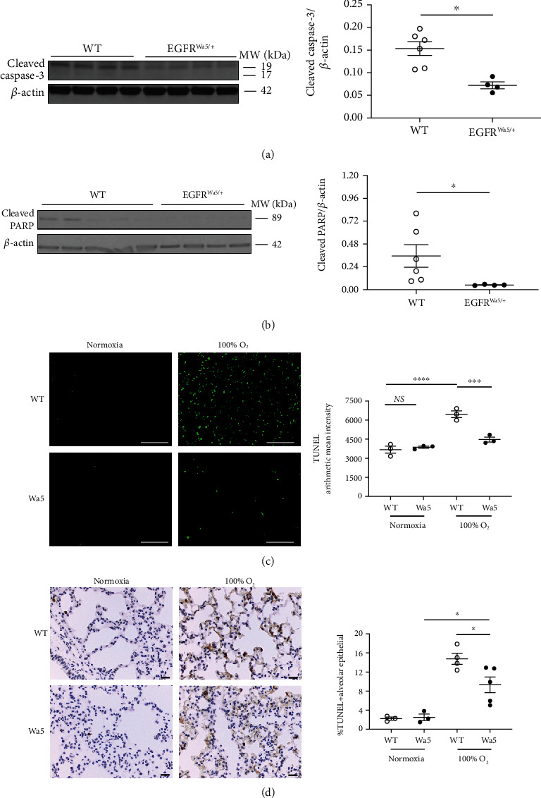
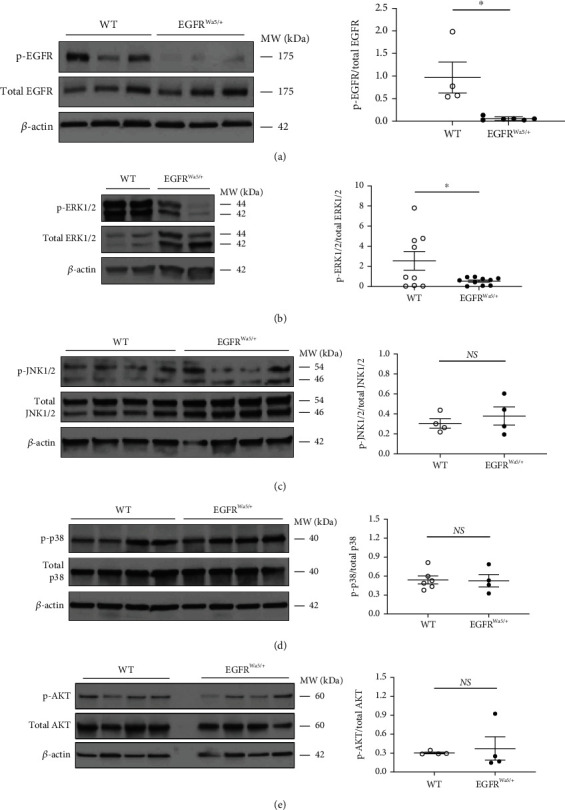
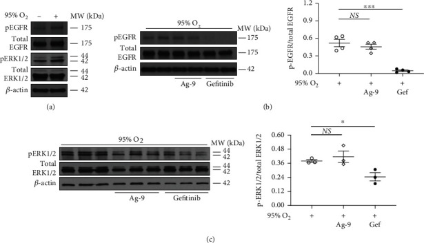
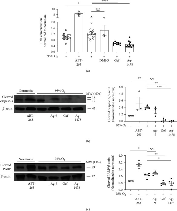
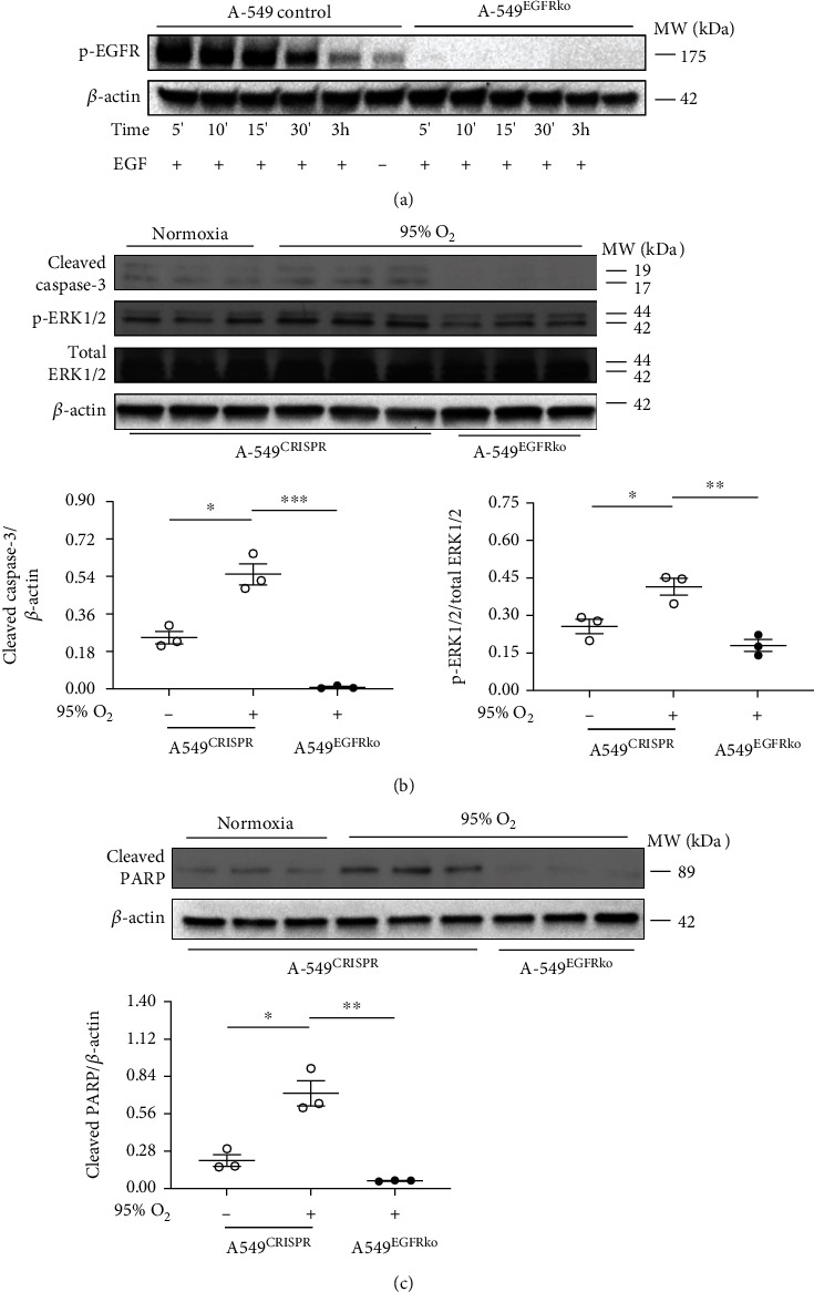
Results: Effects of severe hyperoxia (100% oxygen) were studied in mice with genetically inhibited EGFR and wild-type littermates. Despite the established role of EGFR in lung repair, EGFR inhibition led to improved survival and reduced acute lung injury, which prompted an investigation into this protective mechanism. Endothelial EGFR genetic knockout did not confer protection. EGFR inhibition led to decreased levels of cleaved caspase-3 and poly (ADP-ribosyl) polymerase (PARP) and decreased terminal dUTP nick end labeling- (TUNEL-) positive staining in alveolar epithelial cells and reduced ERK activation, which suggested reduced apoptosis in vivo. EGFR inhibition decreased hyperoxia (95%)-induced apoptosis and ERK in murine alveolar epithelial cells in vitro, and CRISPR-mediated EGFR deletion reduced hyperoxia-induced apoptosis and ERK in human alveolar epithelial cells in vitro. Innovation. This work defines a protective role of EGFR inhibition to decrease apoptosis in lung injury induced by 100% oxygen. This further characterizes the complex role of EGFR in acute lung injury and outlines a novel hyperoxia-induced cell death pathway that warrants further study.

Conclusion: In conditions of severe hyperoxia (>95% for >24 h), EGFR inhibition led to improved survival, decreased lung injury, and reduced cell death. These findings further elucidate the complex role of EGFR in acute lung injury.


Conflict of interest statement

The authors declare that there is no conflict of interest regarding the publication of this article.

Figures

Figure 1
Figure 1
EGFR inhibition improves survival and reduces acute lung injury in hyperoxia. (a) Effects of severe hyperoxia (100% oxygen) on survival were examined in EGFRWa5/+ mice vs. WT littermates (N = 5-6 mice/group, repeated twice) ∗p < 0.05, log-rank analysis. (b–f) BAL and lungs were analyzed in EGFRWa5/+ and WT mice subjected to hyperoxia for 24 h (N = 5-6 mice/group, repeated once), 48 h (N = 5-6 mice/group, repeated twice), and 72 h (N = 5-6 mice/group, repeated twice). BAL at 48 h: (b) cell count. ∗p < 0.05, Mann-Whitney U test. (c) Cell differential. ∗p < 0.05, Mann-Whitney U test. (d) 24, 48, and 72 h for BAL lactate dehydrogenase (LDH). ∗p < 0.05, Mann-Whitney U test. (e) BAL total protein at 48 h. (f) Histopathology (H&E) of lungs from EGFRWa5/+ and WT mice at 72 h severe hyperoxia (representative of N = 5-6 mice/group, repeated twice, scale bar = 100 μm). BAL: bronchoalveolar lavage; L: lymphocytes; Mϕ: macrophages; N: neutrophils; Wa5: EGFRWa5/+ mice; WT: wild type.
Figure 2
Figure 2
Endothelial-specific EGFR knockout is not protective in hyperoxia-induced lung injury. (a) To determine EGFR expression by pulmonary cell type, publicly available datasets of single-cell RNA sequencing (scRNA seq) on the mouse lung were analyzed as described in the methods. EGFR expression levels for each cell type is shown. (b) Effects of severe hyperoxia (100% oxygen) on survival were examined in EGFREndoKO mice vs. control littermates (N = 5-6 mice/group). (c–e) BAL and lungs were analyzed in EGFREndoKO mice and WT mice subjected to hyperoxia for 48 h (N = 5-6 mice/group) and 72 h (N = 5-6 mice/group). BAL at 48 h: (c) cell count. (d) 48 h and 72 h for BAL lactate dehydrogenase (LDH). (e) BAL total protein at 48 h. BAL: bronchoalveolar lavage; DC: dendritic cell; EGFREndoKO: mice with endothelial-specific knockout of EGFR; ILC: innate lymphoid cell; NK cell: natural killer cell; NKT: natural killer T cell; Tgd: γδ T cell.
Figure 3
Figure 3
EGFR inhibition decreases pulmonary cell death and apoptotic markers in hyperoxia-induced lung injury. (a–d) Effects of severe hyperoxia (100% oxygen) were examined in EGFRWa5/+ and WT mice at 24 h (N = 5-6 mice/group, repeated once), 48 h (N = 5-6 mice/group, repeated twice), and 72 h (N = 5-6 mice/group, repeated twice). (a) Western blot (WB) for cleaved caspase-3 at 48 h in whole lungs. Representative samples run in duplicate are shown (representative of three independent experiments). Quantitative densitometry using β-actin is shown. ∗p < 0.05, Mann-Whitney U test. (b) WB for cleaved PARP at 48 h in the whole lungs. WB of three independent experiments is shown. Quantitative densitometry using β-actin is shown. ∗p < 0.05, Mann-Whitney U test. (c, d) TUNEL on lungs from EGFRWa5/+ and WT mice at 72 h severe hyperoxia (representative of three independent experiments, N = 5-6 mice/group, scale bar = 100 μm). (c) TUNEL immunofluorescent staining. Quantification of TUNEL arithmetic mean intensity is shown. ∗∗∗p < 0.0005, ∗∗∗∗p < 0.0001, 2-way ANOVA. (d) TUNEL staining with hematoxylin counterstain. Quantification of TUNEL-positive alveolar epithelial cells expressed as percentage of total number of alveolar epithelial cells is shown. ∗p < 0.05, unpaired t test. PARP: poly (ADP-ribosyl) polymerase; TUNEL: terminal dUTP nick end labeling; Wa5: EGFRWa5/+ mice; WT: wild type.
Figure 4
Figure 4
EGFR is activated in hyperoxia and modulates ERK, but not JNK, p38, and AKT in vivo. Effects of severe hyperoxia (100% oxygen) were examined in lungs from EGFRWa5/+ and WT mice at 24 (N = 5-6 mice/group, repeated once), 48 (N = 5-6 mice/group, repeated twice), and 72 h (N = 5-6 mice/group, repeated twice). (a) Western blot (WB) for EGFR (p- and total) at 48 h. ∗p < 0.05, Mann-Whitney U test. (b) WB for ERK1/2 (p- and total) at 24 h. ∗p < 0.05, unpaired t test. (c) WB for JNK1/2 (p- and total) at 48 h. (d) WB for p38 (p- and total) at 48 h. (e) WB for p-AKT (p- and total) at 72 h. WB of two independent experiments (24 h) and three independent experiments (48 h and 72 h) is shown. Densitometry using total fraction is shown. ERK1/2: extracellular signal-regulated kinase 1 and 2; JNK1/2: c-Jun N-terminal kinase 1 and 2; p38: protein 38; WT: wild type.
Figure 5
Figure 5
Hyperoxia activates ERK via EGFR in murine alveolar epithelial cells. Effects of severe hyperoxia (95% oxygen) were examined in MLE12 cells treated with EGFR-selective tyrosine kinase inhibitor gefitinib and compared with normoxia (repeated twice). Ag-9 was used as a negative control. Western blot (WB) for (a) EGFR (p- and total) and ERK1/2 (p- and total) at 60 minutes. Representative sample is shown (representative of three independent experiments). (b) EGFR (p- and total) at 60 minutes. WB of three independent experiments is shown. Quantitative densitometry of 2 representative samples run in duplicate using total fraction is shown. ∗∗∗p < 0.0005, unpaired t test. (c) ERK1/2 (p- and total) at 48 h. Quantitative densitometry using total fraction is shown. ∗p < 0.05, unpaired t test. ERK1/2: extracellular signal-regulated kinase 1 and 2; Gef: gefitinib; Hyp: hyperoxia; MLE12: murine lung epithelial 12.
Figure 6
Figure 6
EGFR inhibition decreases cell death and apoptotic markers in murine alveolar epithelial cells in severe hyperoxia. Effects of severe hyperoxia (95% oxygen) were examined in MLE12 cells treated with EGFR-selective tyrosine kinase inhibitors gefitinib and Ag-1478 and compared with normoxia controls (repeated twice). ABT-263, an apoptosis inducer, was used as positive control. DMSO and Ag-9 were used as negative controls. (a) Lactate dehydrogenase (LDH) at 72 h. Representative of three independent experiments. LDH concentration values for each experiment were normalized to the average normoxia LDH concentration for that experiment. ∗p < 0.05, ∗∗∗∗p < 0.001 unpaired t test. (b, c) Western blot (WB) for (b) cleaved caspase-3 and (c) cleaved PARP at 48 h. WB of three independent experiments is shown. Quantitative densitometry using β-actin is shown. Quantitative densitometry values for each WB were normalized to the normoxia densitometry value (i.e., control value) for that WB. ∗p < 0.05, ∗∗p < 0.005, and ∗∗∗p < 0.0005; unpaired t test. Gef: gefitinib; Hyp: hyperoxia; PARP: poly (ADP-ribosyl) polymerase.
Figure 7
Figure 7
EGFR deletion via CRISPR leads to decreased apoptotic markers and reduced ERK activation in human alveolar epithelial cells in hyperoxia. (a) To confirm EGFR deletion, A-549 cells containing CRISPR-mediated EGFR deletion (A-549EGFRko) and relevant controls (A-549 (control)) were stimulated with EGF. Western blot (WB) for p-EGFR is shown. (b, c) Effects of severe hyperoxia (95% oxygen) were examined in A-549EGFRko and relevant controls (A-549 (control) and cells containing empty CRISPR vector (A-549CRISPR)) and compared with normoxia. (b) WB for cleaved caspase-3 and ERK1/2 (p- and total) at 48 h. Quantitative densitometry using β-actin for caspase-3 and total-ERK1/2 for p-ERK is shown. ∗p < 0.05, ∗∗p < 0.005, and ∗∗∗p < 0.0005; unpaired t test. (c) WB for cleaved PARP at 48 h. Quantitative densitometry using β-actin is shown. ∗p < 0.05, ∗∗p < 0.005; unpaired t test. Hyp: hyperoxia; PARP: poly (ADP-ribosyl) polymerase.

Similar articles

Cited by

References

    1. Thompson B. T., Chambers R. C., Liu K. D. Acute respiratory distress syndrome. The New England Journal of Medicine . 2017;377(6):562–572. doi: 10.1056/NEJMra1608077. - DOI - PubMed
    1. Budinger G. R. S., Mutlu G. M. Balancing the risks and benefits of oxygen therapy in critically III adults. Chest . 2013;143(4):1151–1162. doi: 10.1378/chest.12-1215. - DOI - PMC - PubMed
    1. Barber R. E., Hamilton W. K. Oxygen toxicity in man. The New England Journal of Medicine . 1970;283(27):1478–1484. doi: 10.1056/NEJM197012312832702. - DOI - PubMed
    1. Clark J. M., Lambertsen C. J. Pulmonary oxygen toxicity: a review. Pharmacological Reviews . 1971;23(2):37–133. - PubMed
    1. Freeman B. A., Crapo J. D. Hyperoxia increases oxygen radical production in rat lungs and lung mitochondria. The Journal of Biological Chemistry . 1981;256(21):10986–10992. doi: 10.1016/S0021-9258(19)68544-3. - DOI - PubMed