Review
. 2023 Feb;306(2):e211658.
doi: 10.1148/radiol.211658. Epub 2022 Oct 4.

MRI Evaluation of Uterine Masses for Risk of Leiomyosarcoma: A Consensus Statement

Affiliations
Review

MRI Evaluation of Uterine Masses for Risk of Leiomyosarcoma: A Consensus Statement

Nicole Hindman et al. Radiology. 2023 Feb.

Abstract

Laparoscopic myomectomy, a common gynecologic operation in premenopausal women, has become heavily regulated since 2014 following the dissemination of unsuspected uterine leiomyosarcoma (LMS) throughout the pelvis of a physician treated for symptomatic leiomyoma. Research since that time suggests a higher prevalence than previously suspected of uterine LMS in resected masses presumed to represent leiomyoma, as high as one in 770 women (0.13%). Though rare, the dissemination of an aggressive malignant neoplasm due to noncontained electromechanical morcellation in laparoscopic myomectomy is a devastating outcome. Gynecologic surgeons' desire for an evidence-based, noninvasive evaluation for LMS is driven by a clear need to avoid such harms while maintaining the availability of minimally invasive surgery for symptomatic leiomyoma. Laparoscopic gynecologists could rely upon the distinction of higher-risk uterine masses preoperatively to plan oncologic surgery (ie, potential hysterectomy) for patients with elevated risk for LMS and, conversely, to safely offer women with no or minimal indicators of elevated risk the fertility-preserving laparoscopic myomectomy. MRI evaluation for LMS may potentially serve this purpose in symptomatic women with leiomyomas. This evidence review and consensus statement defines imaging and disease-related terms to allow more uniform and reliable interpretation and identifies the highest priorities for future research on LMS evaluation.


Conflict of interest statement

Disclosures of conflicts of interest: N.H. Member of the Radiology Editorial Board. S.K. Grants from the National Cancer Institute (National Institutes of Health) and the Doris Duke Foundation; royalties from Wolters Kluwer; chair of the Incidental Findings Steering Committee of the American College of Radiology and specialty chair of Gynecologic & Obstetrical Imaging, Appropriateness Criteria, of the American College of Radiology; member of the Radiology Editorial Board. L.F. Speaker fees from GE Healthcare; travel and meeting expenses from Guerbet. Y.L. Supported by the National Institutes of Health/National Cancer Institute Cancer Center Support Grant (P30 CA008748); grant to institution from the National Cancer Institute; stock or stock options in Y-mAbs Therapeutics; consultant for Calyx Clinical Trial Solutions. S.N. No relevant relationships. C.R. No relevant relationships. E.S. No relevant relationships. J.Q.H. No relevant relationships. S.A. No relevant relationships.

Figures

None
Graphical abstract
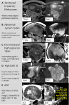
(A–E) MRI scans depict features with a strong association with
leiomyosarcoma (LMS). All images are in the axial plane, except for the
second column of row C, which is a sagittal T2-weighted image. The short
arrow in the left panel of D indicates signal intensity in the LMS, and the
long arrow shows signal intensity in the endometrium. In the right panel of
D, the arrow shows signal intensity in a lymph node. Apparent diffusion
coefficients (ADCs) are expressed in square millimeters per second ×
10−3. b1000 = b value of 1000 sec/mm2, Circumf = circumference, Dia =
diameter, DWI = diffusion-weighted imaging, Max = maximum, Min = minimum,
ROI = region of interest, SI = signal intensity, T2WI = T2-weighted
imaging.
Figure 1:
(A–E) MRI scans depict features with a strong association with leiomyosarcoma (LMS). All images are in the axial plane, except for the second column of row C, which is a sagittal T2-weighted image. The short arrow in the left panel of D indicates signal intensity in the LMS, and the long arrow shows signal intensity in the endometrium. In the right panel of D, the arrow shows signal intensity in a lymph node. Apparent diffusion coefficients (ADCs) are expressed in square millimeters per second × 10−3. b1000 = b value of 1000 sec/mm2, Circumf = circumference, Dia = diameter, DWI = diffusion-weighted imaging, Max = maximum, Min = minimum, ROI = region of interest, SI = signal intensity, T2WI = T2-weighted imaging.
Imaging depiction of consensus definitions of margins of uterine
masses. All images are axial T2-weighted images (T2WI).
Figure 2:
Imaging depiction of consensus definitions of margins of uterine masses. All images are axial T2-weighted images (T2WI).
Flowchart for atypical uterine mass evaluation at MRI. * =
Likely benign has a low likelihood of malignancy; however, given the
conflicting data in the literature for the consensus on which absolute
threshold of the apparent diffusion coefficient (ADC) to use for capturing
all leiomyosarcoma, it is still recommended that masses that fall under this
category be evaluated with a multidisciplinary team for management, with
consideration for open myomectomy. ** = Suspicious for
malignancy indicates that many of the mass lesions in this category are
malignant; however, cellular leiomyomas and smooth muscle tumors of unknown
malignant potential occasionally fall into this category. These masses
should be managed with a multidisciplinary team approach, as described in
Figure 6. DWI = diffusion-weighted imaging.
Figure 3:
Flowchart for atypical uterine mass evaluation at MRI. * = Likely benign has a low likelihood of malignancy; however, given the conflicting data in the literature for the consensus on which absolute threshold of the apparent diffusion coefficient (ADC) to use for capturing all leiomyosarcoma, it is still recommended that masses that fall under this category be evaluated with a multidisciplinary team for management, with consideration for open myomectomy. ** = Suspicious for malignancy indicates that many of the mass lesions in this category are malignant; however, cellular leiomyomas and smooth muscle tumors of unknown malignant potential occasionally fall into this category. These masses should be managed with a multidisciplinary team approach, as described in Figure 6. DWI = diffusion-weighted imaging.
Atypical uterine mass evaluation for leiomyosarcoma (LMS) and its
mimickers: application of the atypical uterine mass flowchart. This figure
demonstrates the real-time application of the Figure 3 flowchart, beginning
with assessment for uterine mass enhancement in column 2. As the columns
progress to the right, the various features in the top row are applied. If
the mass meets criteria for further analysis, an arrow is seen in the box,
indicating that the next feature be applied. Once the mass is determined to
fit benign criteria, a stop symbol is shown and subsequent sequences to the
right are grayed out, indicating that these features should not be
evaluated. Masses characterized as benign according to this imaging
flowchart analysis do not warrant additional work-up from an imaging
perspective. Clinical treatment of such masses can proceed along standard
gynecologic management of benign leiomyomas. All images were acquired in the
axial plane, except for the T2-weighted image in the second row, which was
acquired in the coronal plane. Apparent diffusion coefficient (ADC) is
expressed in square millimeters per second × 10−3. b1000 = b
value of 1000 sec/mm2, CE = contrast-enhanced, DWI = diffusion-weighted
imaging, endo = endometrium, int. = intermediate, STUMP = smooth muscle
tumor of unknown malignant potential, T2WI = T2-weighed imaging.
Figure 4:
Atypical uterine mass evaluation for leiomyosarcoma (LMS) and its mimickers: application of the atypical uterine mass flowchart. This figure demonstrates the real-time application of the Figure 3 flowchart, beginning with assessment for uterine mass enhancement in column 2. As the columns progress to the right, the various features in the top row are applied. If the mass meets criteria for further analysis, an arrow is seen in the box, indicating that the next feature be applied. Once the mass is determined to fit benign criteria, a stop symbol is shown and subsequent sequences to the right are grayed out, indicating that these features should not be evaluated. Masses characterized as benign according to this imaging flowchart analysis do not warrant additional work-up from an imaging perspective. Clinical treatment of such masses can proceed along standard gynecologic management of benign leiomyomas. All images were acquired in the axial plane, except for the T2-weighted image in the second row, which was acquired in the coronal plane. Apparent diffusion coefficient (ADC) is expressed in square millimeters per second × 10−3. b1000 = b value of 1000 sec/mm2, CE = contrast-enhanced, DWI = diffusion-weighted imaging, endo = endometrium, int. = intermediate, STUMP = smooth muscle tumor of unknown malignant potential, T2WI = T2-weighed imaging.
Difficult atypical uterine mass lesion evaluation using the atypical
uterine mass flowchart. This figure demonstrates the real-time application
of the Figure 3 flowchart, beginning with assessment for uterine mass
enhancement in column 2. As the columns progress to the right, the various
features in the top row are applied. If the mass meets criteria for further
analysis, an arrow is seen in the box, indicating that the next feature be
applied. Once the mass is determined to fit benign criteria, a stop symbol
is shown and subsequent sequences to the right are grayed out, indicating
that these features should not be evaluated. Masses characterized as benign
according to this imaging flowchart analysis do not warrant additional
work-up from an imaging perspective. Clinical treatment of such masses can
proceed along standard gynecologic management of benign leiomyomas. Note the
important role of the diffusion-weighted imaging (DWI) sequence with a b
value of 1000 sec/mm2 (b1000) for distinguishing between leiomyosarcoma
(LMS) and benign leiomyoma subtypes, a key feature initially described by
Abdel Wahab et al (54). All images are in the axial plane. Apparent
diffusion coefficient (ADC) is expressed in square millimeters per second
× 10−3. CE = contrast-enhanced, endo = endometrium, int. =
intermediate, LN = lymph node, STUMP = smooth muscle tumor of unknown
malignant potential, T2WI = T2 weighed imaging.
Figure 5:
Difficult atypical uterine mass lesion evaluation using the atypical uterine mass flowchart. This figure demonstrates the real-time application of the Figure 3 flowchart, beginning with assessment for uterine mass enhancement in column 2. As the columns progress to the right, the various features in the top row are applied. If the mass meets criteria for further analysis, an arrow is seen in the box, indicating that the next feature be applied. Once the mass is determined to fit benign criteria, a stop symbol is shown and subsequent sequences to the right are grayed out, indicating that these features should not be evaluated. Masses characterized as benign according to this imaging flowchart analysis do not warrant additional work-up from an imaging perspective. Clinical treatment of such masses can proceed along standard gynecologic management of benign leiomyomas. Note the important role of the diffusion-weighted imaging (DWI) sequence with a b value of 1000 sec/mm2 (b1000) for distinguishing between leiomyosarcoma (LMS) and benign leiomyoma subtypes, a key feature initially described by Abdel Wahab et al (54). All images are in the axial plane. Apparent diffusion coefficient (ADC) is expressed in square millimeters per second × 10−3. CE = contrast-enhanced, endo = endometrium, int. = intermediate, LN = lymph node, STUMP = smooth muscle tumor of unknown malignant potential, T2WI = T2 weighed imaging.
Consensus flowchart of the generalized management algorithm for the
atypical uterine mass visualized with MRI. This management algorithm provides
general guidance for multidisciplinary teams using MRI for leiomyosarcoma (LMS)
screening for patients in whom any level of suspicion of LMS is raised at MRI.
After cervical cancer is excluded by means of Papanicolaou (Pap) smear and
endometrial cancer by means of endometrial biopsy, the patient who wishes to
preserve her uterus is offered a two-step operative procedure. Two-step means
that the patient will potentially have two operations. The first operation is an
open myomectomy, with resection of the uterine mass. Because frozen-section
analysis is only accurate in up to 88% of patients with uterine masses, a
definitive pathologic result is obtained, which can take up to a week. After the
final pathologic result is available, if the patient has a malignant neoplasm
like LMS, then a definitive hysterectomy and/or lymphadenectomy is performed. If
the final pathologic result is benign, then the patient is reassured, her
fertility is preserved, and no further operation is required.
Figure 6:
Consensus flowchart of the generalized management algorithm for the atypical uterine mass visualized with MRI. This management algorithm provides general guidance for multidisciplinary teams using MRI for leiomyosarcoma (LMS) screening for patients in whom any level of suspicion of LMS is raised at MRI. After cervical cancer is excluded by means of Papanicolaou (Pap) smear and endometrial cancer by means of endometrial biopsy, the patient who wishes to preserve her uterus is offered a two-step operative procedure. Two-step means that the patient will potentially have two operations. The first operation is an open myomectomy, with resection of the uterine mass. Because frozen-section analysis is only accurate in up to 88% of patients with uterine masses, a definitive pathologic result is obtained, which can take up to a week. After the final pathologic result is available, if the patient has a malignant neoplasm like LMS, then a definitive hysterectomy and/or lymphadenectomy is performed. If the final pathologic result is benign, then the patient is reassured, her fertility is preserved, and no further operation is required.

References

    1. Baird DD , Dunson DB , Hill MC , Cousins D , Schectman JM . High cumulative incidence of uterine leiomyoma in black and white women: ultrasound evidence . Am J Obstet Gynecol 2003. ; 188 ( 1 ): 100 – 107 . - PubMed
    1. Stewart EA , Cookson CL , Gandolfo RA , Schulze-Rath R . Epidemiology of uterine fibroids: a systematic review . BJOG 2017. ; 124 ( 10 ): 1501 – 1512 . - PubMed
    1. Donnez J , Dolmans MM . Uterine fibroid management: from the present to the future . Hum Reprod Update 2016. ; 22 ( 6 ): 665 – 686 . - PMC - PubMed
    1. Laparoscopic Uterine Power Morcellation in Hysterectomy and Myomectomy: FDA Safety Communication . https://wayback.archive-it.org/7993/20170404182209/https:/www.fda.gov/Me.... Published 2014. Accessed August 5, 2020 .
    1. Durand-Réville M , Dufour P , Vinatier D , et al. . Uterine leiomyosarcomas: a surprising pathology. Review of the literature. Six case reports [in French] . J Gynecol Obstet Biol Reprod (Paris) 1996. ; 25 ( 7 ): 710 – 715 . - PubMed

Publication types