. 2022 Nov 8;7(21):e140384.
doi: 10.1172/jci.insight.140384.

IRF4 expression by lung dendritic cells drives acute but not Trm cell-dependent memory Th2 responses

Affiliations

IRF4 expression by lung dendritic cells drives acute but not Trm cell-dependent memory Th2 responses

Daniel F Camacho et al. JCI Insight. .

Abstract

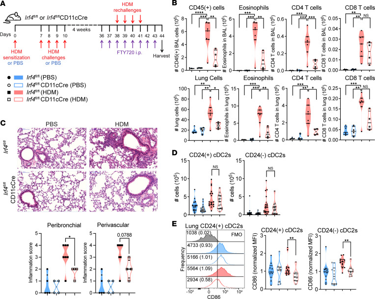
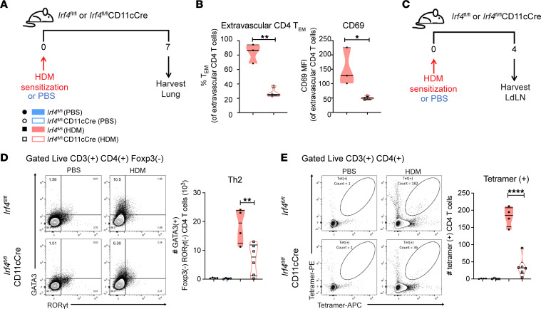
Expression of the transcription factor interferon regulatory factor 4 (IRF4) is required for the development of lung conventional DCs type 2 (cDC2s) that elicit Th2 responses, yet how IRF4 functions in lung cDC2s throughout the acute and memory allergic response is not clear. Here, we used a mouse model that loses IRF4 expression after lung cDC2 development to demonstrate that mice with IRF4-deficient DCs display impaired memory responses to allergen. This defect in the memory response was a direct result of ineffective Th2 induction and impaired recruitment of activated effector T cells to the lung after sensitization. IRF4-deficient DCs demonstrated defects in their migration to the draining lymph node and in T cell priming. Finally, T cells primed by IRF4-competent DCs mediated potent memory responses independently of IRF4-expressing DCs, demonstrating that IRF4-expressing DCs are not necessary during the memory response. Thus, IRF4 controlled a program in mature DCs governing Th2 priming and effector responses, but IRF4-expressing DCs were dispensable during tissue-resident memory T cell-dependent memory responses.

Keywords: Asthma; Dendritic cells; Immunology; Th2 response.


Conflict of interest statement

Conflict of interest: The authors have declared that no conflict of interest exist.

Figures

Figure 1
Figure 1. IRF4-expressing DCs regulate the Trm cell–restricted type 2 inflammatory memory response to HDM rechallenges.
(A) Schematic of the experimental protocol for Trm cell–restricted memory response to HDM. (B) Total cellularity, eosinophils, CD4+ T cells, and CD8+ T cells in the airways (top) or lungs (bottom); n = 20. (C) H&E staining of the lungs confirms that mice with IRF4-deficient DCs are protected from allergic airway inflammation during the memory recall response to HDM; n = 20. Scale bar: 100 μm. (D) Number of CD24+ cDC2s and CD24 cDC2s in the lungs; n = 63. (E) For lung CD24+ cDC2s and CD24 cDC2s, MFI of CD86 normalized to the mean of the PBS-treated Irf4fl/fl group, with representative flow plots; n = 40. Data are (B and C) representative of or (D and E) combined from 3 independent experiments with n ≥ 4 mice per group in each experiment; statistics (B, ordinary 1-way ANOVA with Tukey’s multiple comparisons test; C and D, Mann-Whitney test) were performed in GraphPad Prism. Data are shown as the mean ± SEM (*P < 0.05; **P < 0.01; ***P < 0.001; ****P < 0.0001). Also see Supplemental Figures 2 and 4.
Figure 2
Figure 2. Lungs of mice with IRF4-deficient DCs contain fewer Der p 1–specific Th2rm cells during the memory phase.
(A) Schematic of experimental protocol for resting memory lung analysis. (B) Number of lung Trm cells and the proportion expressing ST2; n = 20. Data are representative of 3 independent experiments with n ≥ 4 mice per group in each experiment. Analysis was performed by ordinary 1-way ANOVA with Tukey’s multiple comparisons test. (C) Gating of antigen-specific T cells and their expression of lineage-defining transcription factors. (D) Graphed flow plots show the number of tetramer+CD4+ Trm cells expressing GATA3 or Foxp3; n = 36. Three independent experiments (n ≥ 5 per group) were statistically significant. Two of these experiments combined are shown. Kruskal-Wallis test (with Dunn’s multiple comparisons test) was performed in GraphPad Prism. Data are shown as the mean ± SEM (*P < 0.05; **P < 0.01). Also see Supplemental Figures 3 and 4.
Figure 3
Figure 3. Mice with IRF4-deficient DCs are unable to adequately recruit Tem cells to the lungs and produce Th2 cells or tetramer+ T cells in the lung draining lymph nodes after HDM sensitization.
(A) Schematic of experimental protocol for analysis of initiation of Th2 responses during sensitization phase in the lung. (B) Proportion of T effector cells of lung extravascular CD4+ T cells, and CD69 expression by extravascular lung T cells; n = 7. (C) Schematic of experimental protocol for analysis of initiation of Th2 responses during sensitization phase in the lung draining lymph nodes (LdLNs). (D) GATA3 and RORγt expression by LdLN T conventional cells on day 4 after HDM sensitization; n = 18. (E) Number of tetramer+ LdLN CD4+ T cells; n = 18. Data are representative of 2 independent experiments with n ≥ 3 mice per group; statistics (B, unpaired t test with Welch’s correction; D and E, ordinary 1-way ANOVA with Tukey’s multiple comparisons test) were performed in GraphPad Prism. Data are shown as the mean ± SEM (*P < 0.05; **P < 0.01; ****P < 0.0001).
Figure 4
Figure 4. IRF4 regulates allergen phagocytosis and migration of CD24+ cDC2s to the lung draining lymph nodes during HDM sensitization.
(A and B) Flow cytometry plots depict fluorescently labeled HDM in CD24+ cDC2s, quantified number of CD24+ cDC2s, the proportion and number that were HDM+, and MFI of HDM in either (A) the lungs (n = 11) or (B) lung draining lymph nodes (LdLNs; n = 10), as 1 LdLN could not be harvested. (C and D) Flow cytometry plots depict fluorescence of processed DQ Red BSA in CD24+ cDC2s, proportion DQ+, and MFI of DQ in either (C) the lungs (n = 10) or (D) LdLN (n = 9). Data are representative of 2 independent experiments with n ≥ 4 mice per group; statistics (unpaired t test with Welch’s correction) were performed in GraphPad Prism. Data are shown as the mean ± SEM (*P < 0.05; ***P < 0.001). Also see Supplemental Figure 5.
Figure 5
Figure 5. Ex vivo–sorted CD24+ cDC2s require IRF4 for robust T cell priming in vitro.
(A) Schematic of experimental protocol for in vivo sensitization to HDM+ OVA, DC sorting, and in vitro coculture with CFSE-labeled T cells from naive OTII mice. (B and C) Number of OTII cells after culture, percentage undivided, division index, proliferation index, and CFSE dilution histograms for (B) in vivo HDM+OVA–sensitized CD24+ cDC2s (n = 20) or (C) those with OVA323–339 peptide added (n = 20). (B and C) Data are representative of 2 independent experiments with n ≥ 4 wells per group; statistics (unpaired t test with Welch’s correction) were performed in GraphPad Prism. (D) IL-33 and IL-10 expression by qPCR of sorted lung cDC2s after in vivo HDM sensitization; n = 6. Data represent 1 experiment with n = 3 mice per group; statistics (unpaired t test) were performed in GraphPad Prism. Data are shown as the mean ± SEM (*P < 0.05; **P < 0.01; ****P < 0.0001). Also see Supplemental Figure 6.
Figure 6
Figure 6. IRF4 expression in DCs is not required for CD4+ Trm cell maintenance or recall responses.
(A) Ly5.1 mice were sensitized and challenged with HDM. Lung Tem cells were adoptively transferred to Irf4fl/fl or Irf4fl/flCD11cCre mice and pulled to the lungs with intratracheal rIL-33 in the “Cells + rIL-33” group. Control groups received no cells and either rIL-33 alone or no treatment at all. Lungs were harvested after 4 weeks. (B) Quantified number of donor-derived Trm cells, donor-derived tetramer+ Trm cells, and ST2+ donor-derived Trm cells. Data represent 1 experiment with n ≥ 4 mice per group and a total of n = 21; statistics (Mann-Whitney test) were performed in GraphPad Prism. Data are shown as the mean ± SEM. (C) As in A, but followed by HDM challenges during FTY720 treatment. (D) Number of total cells, eosinophils, CD4+ T cells, and donor CD4+ T cells in the airways normalized to the mean of the Irf4fl/fl group receiving donor cells. Data represent 4 combined experiments with n ≥ 4 mice per group and a total of n = 52; statistics (Mann-Whitney test) were performed in GraphPad Prism. Data are shown as the mean ± SEM. Also see Supplemental Figure 6.

References

    1. Nguyen QP, et al. Origins of CD4+ circulating and tissue-resident memory T-cells. Immunology. 2019;CD4(1):3–12. - PMC - PubMed
    1. Zheng MZM, Wakim LM. Tissue resident memory T cells in the respiratory tract. Mucosal Immunol. 2021;15(3):379–388. - PMC - PubMed
    1. Mackay LK, et al. Long-lived epithelial immunity by tissue-resident memory T (TRM) cells in the absence of persisting local antigen presentation. Proc Natl Acad Sci U S A. 2012;109(18):7037–7042. doi: 10.1073/pnas.1202288109. - DOI - PMC - PubMed
    1. Park SL, et al. Local proliferation maintains a stable pool of tissue-resident memory T cells after antiviral recall responses. Nat Immunol. 2018;19(2):183–191. doi: 10.1038/s41590-017-0027-5. - DOI - PubMed
    1. Jiang X, et al. Skin infection generates non-migratory memory CD8+ T(RM) cells providing global skin immunity. Nature. 2012;483(7388):227–231. doi: 10.1038/nature10851. - DOI - PMC - PubMed

Publication types